Szema Anthony M, Forsyth Edward, Ying Benjamin, Hamidi Sayyed A, Chen John J, Hwang Sonya, Li Jonathan C, Sabatini Dwyer Debra, Ramiro-Diaz Juan M, Giermakowska Wieslawa, Gonzalez Bosc Laura V
Stony Brook University, Department of Technology and Society, College of Engineering and Applied Sciences, Stony Brook, NY, United States of America.
The Stony Brook Medicine SUNY at Stony Brook Internal Medicine Residency Program at John T. Mather Memorial Hospital, Port Jefferson, NY, United States of America.
PLoS One. 2017 Jan 26;12(1):e0170606. doi: 10.1371/journal.pone.0170606. eCollection 2017.
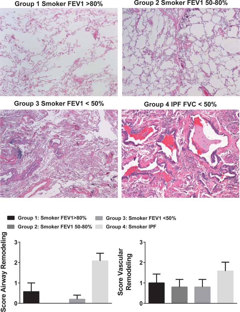
Idiopathic pulmonary fibrosis (IPF) and chronic obstructive pulmonary disease (COPD) are both debilitating lung diseases which can lead to hypoxemia and pulmonary hypertension (PH). Nuclear Factor of Activated T-cells (NFAT) is a transcription factor implicated in the etiology of vascular remodeling in hypoxic PH. We have previously shown that mice lacking the ability to generate Vasoactive Intestinal Peptide (VIP) develop spontaneous PH, pulmonary arterial remodeling and lung inflammation. Inhibition of NFAT attenuated PH in these mice suggesting a connection between NFAT and VIP. To test the hypotheses that: 1) VIP inhibits NFAT isoform c3 (NFATc3) activity in pulmonary vascular smooth muscle cells; 2) lung NFATc3 activation is associated with disease severity in IPF and COPD patients, and 3) VIP and NFATc3 expression correlate in lung tissue from IPF and COPD patients. NFAT activity was determined in isolated pulmonary arteries from NFAT-luciferase reporter mice. The % of nuclei with NFAT nuclear accumulation was determined in primary human pulmonary artery smooth muscle cell (PASMC) cultures; in lung airway epithelia and smooth muscle and pulmonary endothelia and smooth muscle from IPF and COPD patients; and in PASMC from mouse lung sections by fluorescence microscopy. Both NFAT and VIP mRNA levels were measured in lungs from IPF and COPD patients. Empirical strategies applied to test hypotheses regarding VIP, NFATc3 expression and activity, and disease type and severity. This study shows a significant negative correlation between NFAT isoform c3 protein expression levels in PASMC, activity of NFATc3 in pulmonary endothelial cells, expression and activity of NFATc3 in bronchial epithelial cells and lung function in IPF patients, supporting the concept that NFATc3 is activated in the early stages of IPF. We further show that there is a significant positive correlation between NFATc3 mRNA expression and VIP RNA expression only in lungs from IPF patients. In addition, we found that VIP inhibits NFAT nuclear translocation in primary human pulmonary artery smooth muscle cells (PASMC). Early activation of NFATc3 in IPF patients may contribute to disease progression and the increase in VIP expression could be a protective compensatory mechanism.
特发性肺纤维化(IPF)和慢性阻塞性肺疾病(COPD)都是使人衰弱的肺部疾病,可导致低氧血症和肺动脉高压(PH)。活化T细胞核因子(NFAT)是一种转录因子,与低氧性PH的血管重塑病因有关。我们之前已经表明,缺乏产生血管活性肠肽(VIP)能力的小鼠会发生自发性PH、肺动脉重塑和肺部炎症。抑制NFAT可减轻这些小鼠的PH,提示NFAT与VIP之间存在联系。为了验证以下假设:1)VIP抑制肺血管平滑肌细胞中NFAT异构体c3(NFATc3)的活性;2)肺组织中NFATc3的激活与IPF和COPD患者的疾病严重程度相关;3)IPF和COPD患者肺组织中VIP和NFATc3的表达相关。在来自NFAT荧光素酶报告基因小鼠的分离肺动脉中测定NFAT活性。通过荧光显微镜在原代人肺动脉平滑肌细胞(PASMC)培养物中;在IPF和COPD患者的肺气道上皮、平滑肌以及肺内皮和平滑肌中;以及在小鼠肺切片的PASMC中,测定具有NFAT核积累的细胞核百分比。在IPF和COPD患者的肺组织中测量NFAT和VIP的mRNA水平。应用实证策略来检验关于VIP、NFATc3表达和活性以及疾病类型和严重程度的假设。本研究表明,IPF患者PASMC中NFAT异构体c3蛋白表达水平、肺内皮细胞中NFATc3活性、支气管上皮细胞中NFATc3的表达和活性与肺功能之间存在显著负相关,支持NFATc3在IPF早期被激活的概念。我们进一步表明,仅在IPF患者的肺组织中,NFATc3 mRNA表达与VIP RNA表达之间存在显著正相关。此外,我们发现VIP抑制原代人肺动脉平滑肌细胞(PASMC)中NFAT的核转位。IPF患者中NFATc3的早期激活可能有助于疾病进展,而VIP表达的增加可能是一种保护性代偿机制。