• 文献检索
  • 文档翻译
  • 深度研究
  • 学术资讯
  • Suppr Zotero 插件Zotero 插件
  • 邀请有礼
  • 套餐&价格
  • 历史记录
应用&插件
Suppr Zotero 插件Zotero 插件浏览器插件Mac 客户端Windows 客户端微信小程序
定价
高级版会员购买积分包购买API积分包
服务
文献检索文档翻译深度研究API 文档MCP 服务
关于我们
关于 Suppr公司介绍联系我们用户协议隐私条款
关注我们

Suppr 超能文献

核心技术专利:CN118964589B侵权必究
粤ICP备2023148730 号-1Suppr @ 2026

文献检索

告别复杂PubMed语法,用中文像聊天一样搜索,搜遍4000万医学文献。AI智能推荐,让科研检索更轻松。

立即免费搜索

文件翻译

保留排版,准确专业,支持PDF/Word/PPT等文件格式,支持 12+语言互译。

免费翻译文档

深度研究

AI帮你快速写综述,25分钟生成高质量综述,智能提取关键信息,辅助科研写作。

立即免费体验

基于口腔鳞状细胞癌(OCSCC)颈部淋巴结分区相关性分析的临床靶区勾画

The clinical target volume delineation based on the correlation analysis of neck node levels in oral cavity squamous cell carcinoma (OCSCC).

作者信息

Jiang Chaoyang, Li Jiawei, Peng Haibo, Zhou Daijun, Xu Chuan, Zhang Ling, Wang Juan, Liu Bisheng, Li Dong

机构信息

Department of Oncology, The General Hospital of Western Theater Command, Chengdu, China.

Department of Oncology, The Affiliated Hospital of Southwest Medical University, Luzhou, China.

出版信息

BMC Cancer. 2024 Dec 18;24(1):1530. doi: 10.1186/s12885-024-13251-0.

DOI:10.1186/s12885-024-13251-0
PMID:39696109
原文链接:https://pmc.ncbi.nlm.nih.gov/articles/PMC11657834/
Abstract

BACKGROUND

The delineation of clinical target volume (CTV) base on the correlation analysis between neck node levels of OCSCC has not been reported in detail. This study analyzes the correlations between the neck node levels in 208 cases of OCSCC, and aims to provide preliminary reference for the CTV delineation in OCSCC patients.

MATERIALS AND METHODS

The records of 208 OCSCC patients were retrospectively analyzed. The neck node levels were evaluated according to the 2013 updated guidelines. The Chi-square test and logistic regression model were used to analyze the correlation of each level.

RESULTS

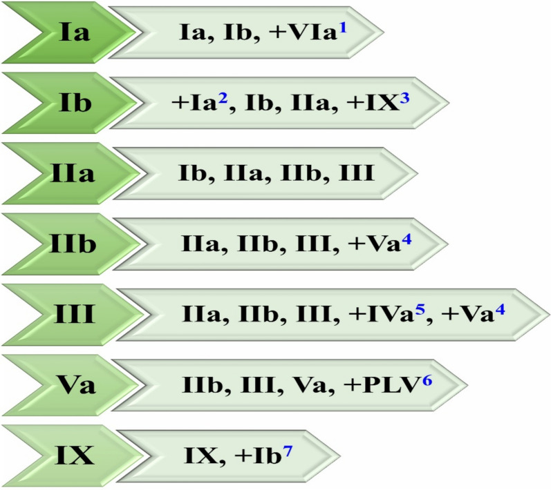
The most common involved level in OCSCC is Ib (45.7%) and IIa (40.4%). Correlation analysis of OCSCC showed that nodal spread in level Ia is related to levels Ib and VIa. Level Ib is related to levels Ia, IIa, IX. Level IIa is related to levels Ib, IIb, III. Level IIb is related to levels IIa, III, Va. Level III is related to levels IIa, IIb, Va. Level Va is related to levels IIb and III. Level VIa is related to level Ia and T stage. Level IX is only related to level Ib. T stage is only related to level VIa. All the above P values are < 0.05.

CONCLUSIONS

The related levels can be considered as high-risk region (HRR) and the unrelated levels as low-risk region (LRR) in the radiotherapy of OCSCC patients. This study can make the CTV delineation more individualized and accurate.

摘要

背景

基于口腔鳞状细胞癌(OCSCC)颈部淋巴结分区相关性分析来勾画临床靶区(CTV)的研究尚未详细报道。本研究分析208例OCSCC患者颈部淋巴结分区的相关性,旨在为OCSCC患者CTV的勾画提供初步参考。

材料与方法

回顾性分析208例OCSCC患者的病历资料。根据2013年更新指南评估颈部淋巴结分区。采用卡方检验和逻辑回归模型分析各分区的相关性。

结果

OCSCC最常累及的分区是Ib(45.7%)和IIa(40.4%)。OCSCC的相关性分析显示,Ia区的淋巴结转移与Ib区和VIa区相关。Ib区与Ia区、IIa区、IX区相关。IIa区与Ib区、IIb区、III区相关。IIb区与IIa区、III区、Va区相关。III区与IIa区、IIb区、Va区相关。Va区与IIb区和III区相关。VIa区与Ia区和T分期相关。IX区仅与Ib区相关。T分期仅与VIa区相关。上述所有P值均<0.05。

结论

在OCSCC患者放疗中,可将相关分区视为高风险区域(HRR),不相关分区视为低风险区域(LRR)。本研究可使CTV的勾画更具个体化和准确性。

https://cdn.ncbi.nlm.nih.gov/pmc/blobs/b0a4/11657834/029932cef34e/12885_2024_13251_Fig2_HTML.jpg
https://cdn.ncbi.nlm.nih.gov/pmc/blobs/b0a4/11657834/bdd3056fd2b2/12885_2024_13251_Fig1_HTML.jpg
https://cdn.ncbi.nlm.nih.gov/pmc/blobs/b0a4/11657834/029932cef34e/12885_2024_13251_Fig2_HTML.jpg
https://cdn.ncbi.nlm.nih.gov/pmc/blobs/b0a4/11657834/bdd3056fd2b2/12885_2024_13251_Fig1_HTML.jpg
https://cdn.ncbi.nlm.nih.gov/pmc/blobs/b0a4/11657834/029932cef34e/12885_2024_13251_Fig2_HTML.jpg

相似文献

1
The clinical target volume delineation based on the correlation analysis of neck node levels in oral cavity squamous cell carcinoma (OCSCC).基于口腔鳞状细胞癌(OCSCC)颈部淋巴结分区相关性分析的临床靶区勾画
BMC Cancer. 2024 Dec 18;24(1):1530. doi: 10.1186/s12885-024-13251-0.
2
Correlation analysis of neck node levels in 960 cases of Nasopharyngeal carcinoma (NPC).960例鼻咽癌颈部淋巴结分区的相关性分析
Radiother Oncol. 2021 Aug;161:23-28. doi: 10.1016/j.radonc.2021.05.020. Epub 2021 May 25.
3
Proposals for the delineation of neck clinical target volume for definitive Radiation therapy in patients with oral/ oropharyngeal squamous cell cancer based on lymph node distribution.基于淋巴结分布的口腔/口咽鳞状细胞癌根治性放疗中颈区临床靶区勾画建议。
Radiother Oncol. 2024 Jun;195:110225. doi: 10.1016/j.radonc.2024.110225. Epub 2024 Mar 14.
4
Contribution of FDG-PET in the diagnostic assessment of cervical lymph node metastasis in Oral Cavity Squamous Cell Carcinoma (OCSCC).18F-FDG PET 在口腔鳞状细胞癌(OCSCC)颈淋巴结转移诊断评估中的作用。
J Stomatol Oral Maxillofac Surg. 2023 Dec;124(6S):101659. doi: 10.1016/j.jormas.2023.101659. Epub 2023 Oct 21.
5
Utility of PET-CT in detecting nodal metastasis in cN0 early stage oral cavity squamous cell carcinoma.PET-CT 在检测 cN0 期口腔鳞癌淋巴结转移中的作用。
Oral Oncol. 2018 May;80:89-92. doi: 10.1016/j.oraloncology.2018.04.003. Epub 2018 Apr 6.
6
Assessment of the Rate of Skip Metastasis to Neck Level IV in Patients With Clinically Node-Negative Neck Oral Cavity Squamous Cell Carcinoma: A Systematic Review and Meta-analysis.评估临床颈部淋巴结阴性口腔鳞状细胞癌患者颈部 IV 水平跳跃转移率:系统评价和荟萃分析。
JAMA Otolaryngol Head Neck Surg. 2019 Jun 1;145(6):542-548. doi: 10.1001/jamaoto.2019.0784.
7
Does Sentinel Lymph Node Biopsy Accurately Stage the Clinically Negative Neck in Early Oral Cavity Squamous Cell Carcinoma?前哨淋巴结活检能否准确分期早期口腔鳞癌临床阴性颈部?
J Oral Maxillofac Surg. 2022 Jun;80(6):1134-1142. doi: 10.1016/j.joms.2022.02.006. Epub 2022 Feb 18.
8
[Analysis of computed tomography-based distribution of metastatic cervical nodes in 218 cases of nasopharyngeal carcinoma].[218例鼻咽癌颈淋巴结转移的CT分布分析]
Ai Zheng. 2004 Sep;23(9):1056-9.
9
Patterns of lymph node involvement for oral cavity squamous cell carcinoma.口腔鳞状细胞癌的淋巴结转移模式。
Radiother Oncol. 2024 Nov;200:110474. doi: 10.1016/j.radonc.2024.110474. Epub 2024 Aug 14.
10
Level V Metastases in Node-Positive Oral Squamous Cell Carcinoma: Beyond Level IIA and III.淋巴结阳性口腔鳞状细胞癌的Ⅴ级转移:超越ⅡA和Ⅲ级。
ORL J Otorhinolaryngol Relat Spec. 2024;86(5-6):174-181. doi: 10.1159/000543140. Epub 2024 Dec 16.

本文引用的文献

1
Proposals for the delineation of neck clinical target volume for definitive Radiation therapy in patients with oral/ oropharyngeal squamous cell cancer based on lymph node distribution.基于淋巴结分布的口腔/口咽鳞状细胞癌根治性放疗中颈区临床靶区勾画建议。
Radiother Oncol. 2024 Jun;195:110225. doi: 10.1016/j.radonc.2024.110225. Epub 2024 Mar 14.
2
The boundary of posterior to level V region and the theoretical feasibility of irradiation dose reduction of level Va in nasopharyngeal carcinoma.鼻咽癌V区后部边界及V a区照射剂量降低的理论可行性
Sci Rep. 2024 Jan 28;14(1):2308. doi: 10.1038/s41598-024-52857-z.
3
Postoperative radiotherapy for supraglottic cancer on real-world data: can we reduce dose to lymph node levels?
基于真实世界数据的声门上型癌症术后放疗:我们能否降低淋巴结水平的剂量?
Radiat Oncol. 2023 Feb 22;18(1):35. doi: 10.1186/s13014-023-02228-1.
4
Incidence and impact of skip metastasis in the neck in early oral cancer: Reality or a myth?早期口腔癌中颈部跳跃性转移的发生率和影响:现实还是虚构?
Oral Oncol. 2022 Dec;135:106201. doi: 10.1016/j.oraloncology.2022.106201. Epub 2022 Oct 12.
5
The predictive value of depth of invasion and tumor size on risk of neck node metastasis in squamous cell carcinoma of the oral cavity: A prospective study.口腔鳞状细胞癌中浸润深度和肿瘤大小对颈部淋巴结转移风险的预测价值:一项前瞻性研究。
J Cancer Res Ther. 2022 Jul-Sep;18(4):977-983. doi: 10.4103/jcrt.JCRT_783_20.
6
Correlation analysis of neck node levels in 960 cases of Nasopharyngeal carcinoma (NPC).960例鼻咽癌颈部淋巴结分区的相关性分析
Radiother Oncol. 2021 Aug;161:23-28. doi: 10.1016/j.radonc.2021.05.020. Epub 2021 May 25.
7
Central neck lymph node metastasis in oral squamous cell carcinoma at the floor of mouth.口腔底部口腔鳞状细胞癌的中央颈部淋巴结转移。
BMC Cancer. 2021 Mar 5;21(1):225. doi: 10.1186/s12885-021-07958-7.
8
Cervical lymph node metastasis in squamous cell carcinoma of the buccal mucosa: a retrospective study on pattern of involvement and clinical analysis.颊黏膜鳞癌颈淋巴结转移:一项关于累及模式和临床分析的回顾性研究。
Med Oral Patol Oral Cir Bucal. 2021 Jan 1;26(1):e84-e89. doi: 10.4317/medoral.24016.
9
Head and neck squamous cell carcinoma.头颈部鳞状细胞癌
Nat Rev Dis Primers. 2020 Nov 26;6(1):92. doi: 10.1038/s41572-020-00224-3.
10
Distribution pattern and prognosis of metastatic lymph nodes in cervical posterior to level V in nasopharyngeal carcinoma patients.鼻咽癌患者颈 V 水平后区转移性淋巴结的分布模式与预后。
BMC Cancer. 2020 Jul 17;20(1):667. doi: 10.1186/s12885-020-07146-z.