Zhang Yuefeng, Mou Zuo, Song Wei, He Xiaoqin, Yi Qin, Wang Zhekai, Mao Xietong, Wang Wei, Xu Yangtao, Shen Yang, Ma Peng, Yu Kaihuan
Department of Hepatobiliary Surgery, Renmin Hospital of Wuhan University, Wuhan, China.
The First Clinical College, Wuhan University, Wuhan, China.
J Nanobiotechnology. 2024 Dec 18;22(1):759. doi: 10.1186/s12951-024-03001-6.
Extracellular vesicles (EVs) and extruded nanovesicles (ENVs) are promising nanovesicles (NVs) for drug delivery. However, the application of these NVs is strongly hindered by their short half-life in the circulation. Macrophages (Mφs) in the liver and spleen contribute to the rapid depletion of NVs, but the underlying mechanism is unclear.
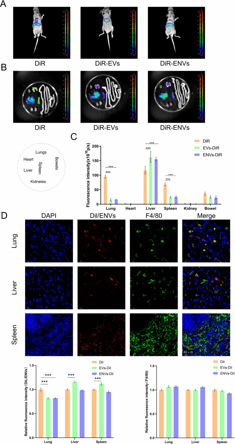
By collecting the supernatant of PANC-1 cells and squeezing PANC-1 cells, EVs and ENVs derived from PANC-1 cells were prepared via ultracentrifugation. NVs were subsequently identified via western blot, particle size measurement, and electron microscopy. The distribution of NVs in mouse bodies was observed with a live animal imaging system. Liver Mφs were extracted and isolated after NVs were administered, and transcriptome profiling was applied to determine differentially expressed genes (DEGs). siRNAs targeting interested genes were designed and synthesized. In vitro experiments, Mφs were transfected with siRNA or treated with the corresponding inhibitor, after which NV uptake was recorded. Doxorubicin (DOX) was encapsulated in ENVs using an ultrasound method. PANC-1 cell-derived tumors were established in nude mice in vivo, inhibitor pretreatment or no treatment was administered before intravenous injection of ENVs-DOX, and the therapeutic efficacy of ENVs-DOX was evaluated.
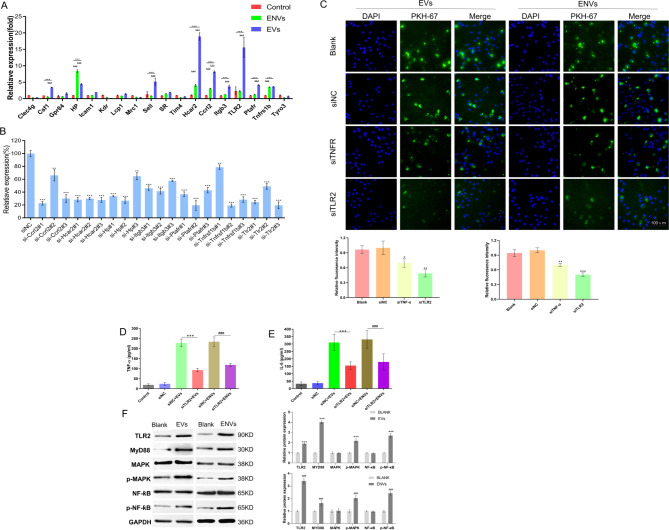
NVs derived from PANC-1 cells were first prepared and identified. After intravenous injection, most NVs were engulfed by Mφs in the liver and spleen. Seven genes of interest were selected via transcriptome sequencing and validated via RT‒PCR. These results confirmed that the TLR2 signaling pathway is responsible for phagocytosis. siTLR2 and its inhibitor sparstolonin B (SpB) significantly inhibited the internalization of NVs by Mφs and downregulated the activity of the TLR2 pathway. The accumulation of ENVs-DOX in the liver was inhibited in vivo by pretreatment with SpB 40 min before intravenous injection, ultimately delaying tumor progression.
The TLR2 pathway plays a crucial role in the sequestration of NVs by Mφs. A novel antiphagocytic strategy in which pretreatment of mice with SpB inhibits the clearance of NVs and prolongs their half-life in vivo, thereby improving delivery efficiency, was identified.
细胞外囊泡(EVs)和挤出纳米囊泡(ENVs)是用于药物递送的有前景的纳米囊泡(NVs)。然而,这些NVs在循环中的半衰期较短,严重阻碍了它们的应用。肝脏和脾脏中的巨噬细胞(Mφs)导致NVs迅速清除,但其潜在机制尚不清楚。
通过收集PANC-1细胞的上清液并挤压PANC-1细胞,经超速离心制备源自PANC-1细胞的EVs和ENVs。随后通过蛋白质免疫印迹、粒度测量和电子显微镜鉴定NVs。用活体动物成像系统观察NVs在小鼠体内的分布。在给予NVs后提取并分离肝脏Mφs,并应用转录组分析来确定差异表达基因(DEGs)。设计并合成靶向感兴趣基因的小干扰RNA(siRNAs)。在体外实验中,用siRNA转染Mφs或用相应抑制剂处理,然后记录NV摄取情况。使用超声方法将阿霉素(DOX)包封在ENVs中。在裸鼠体内建立源自PANC-1细胞的肿瘤,在静脉注射ENVs-DOX之前进行抑制剂预处理或不进行处理,并评估ENVs-DOX的治疗效果。
首次制备并鉴定了源自PANC-1细胞的NVs。静脉注射后,大多数NVs被肝脏和脾脏中的Mφs吞噬。通过转录组测序选择了7个感兴趣的基因,并通过逆转录-聚合酶链反应(RT-PCR)进行验证。这些结果证实Toll样受体2(TLR2)信号通路负责吞噬作用。siTLR2及其抑制剂斯帕索林B(SpB)显著抑制Mφs对NVs的内化,并下调TLR2通路的活性。静脉注射前40分钟用SpB预处理可在体内抑制ENVs-DOX在肝脏中的蓄积,最终延缓肿瘤进展。
TLR2通路在Mφs隔离NVs过程中起关键作用。确定了一种新的抗吞噬策略,即先用SpB预处理小鼠可抑制NVs的清除并延长其在体内的半衰期,从而提高递送效率。