Krefting Research Centre, Institute of Medicine, University of Gothenburg, 40530, Gothenburg, Sweden.
Department of Anesthesiology and Intensive Care Medicine, Institute of Clinical Science, Sahlgrenska Academy, University of Gothenburg, 40530, Gothenburg, Sweden.
Stem Cell Res Ther. 2019 Aug 1;10(1):231. doi: 10.1186/s13287-019-1352-4.
Sepsis remains a source of high mortality in hospitalized patients despite proper antibiotic approaches. Encouragingly, mesenchymal stromal cells (MSCs) and their produced extracellular vesicles (EVs) have been shown to elicit anti-inflammatory effects in multiple inflammatory conditions including sepsis. However, EVs are generally released from mammalian cells in relatively low amounts, and high-yield isolation of EVs is still challenging due to a complicated procedure. To get over these limitations, vesicles very similar to EVs can be produced by serial extrusions of cells, after which they are called nanovesicles (NVs). We hypothesized that MSC-derived NVs can attenuate the cytokine storm induced by bacterial outer membrane vesicles (OMVs) in mice, and we aimed to elucidate the mechanism involved.
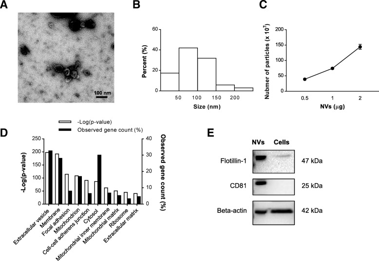
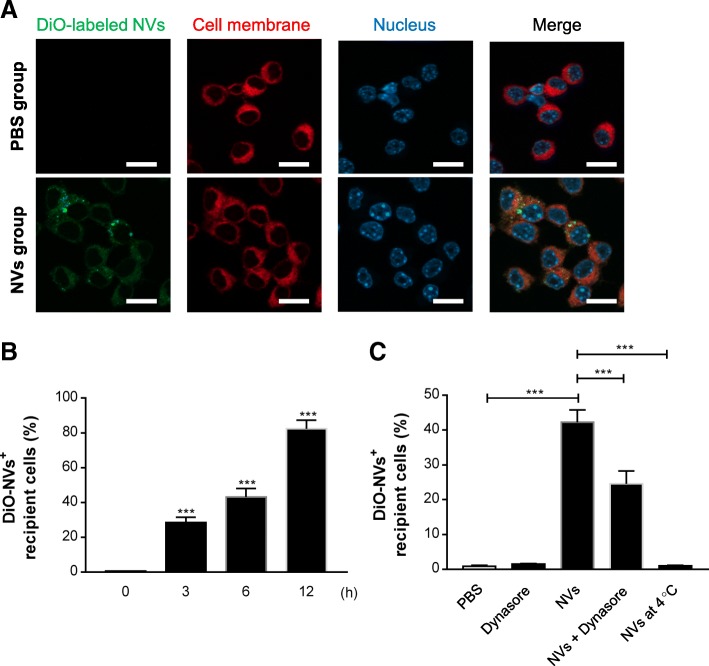
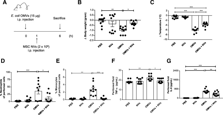
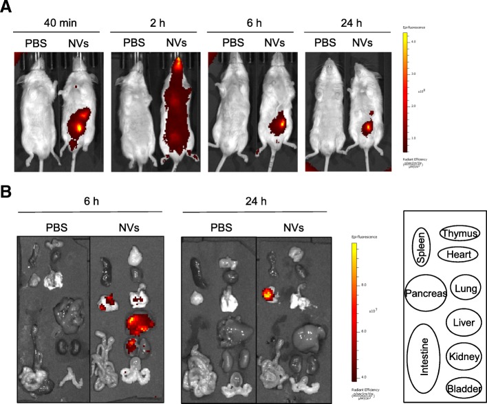
NVs were produced from MSCs by the breakdown of cells through serial extrusions and were subsequently floated in a density gradient. Morphology and the number of NVs were analyzed by transmission electron microscopy and nanoparticle tracking analysis. Mice were intraperitoneally injected with Escherichia coli-derived OMVs to establish sepsis, and then injected with 2 × 10 NVs. Innate inflammation was assessed in peritoneal fluid and blood through investigation of infiltration of cells and cytokine production. The biodistribution of NVs labeled with Cy7 dye was analyzed using near-infrared imaging.
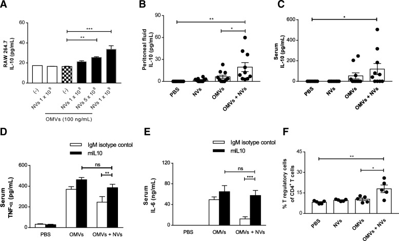
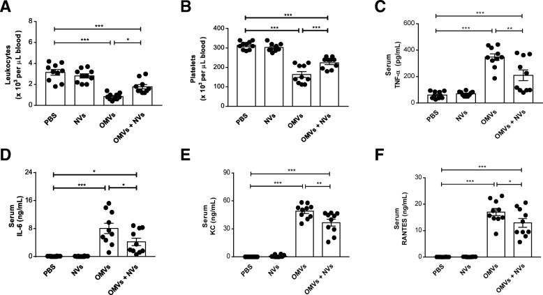
Electron microscopy showed that NVs have a nanometer-size spherical shape and harbor classical EV marker proteins. In mice, NVs inhibited eye exudates and hypothermia, signs of a systemic cytokine storm, induced by intraperitoneal injection of OMVs. Moreover, NVs significantly suppressed cytokine release into the systemic circulation, as well as neutrophil and monocyte infiltration in the peritoneum. The protective effect of NVs was significantly reduced by prior treatment with anti-interleukin (IL)-10 monoclonal antibody. In biodistribution study, NVs spread to the whole mouse body and localized in the lung, liver, and kidney at 6 h.
Taken together, these data indicate that MSC-derived NVs have beneficial effects in a mouse model of sepsis by upregulating the IL-10 production, suggesting that artificial NVs may be novel EV-mimetics clinically applicable to septic patients.
尽管采用了适当的抗生素方法,败血症仍然是住院患者死亡的主要原因。令人鼓舞的是,间充质基质细胞(MSCs)及其产生的细胞外囊泡(EVs)已被证明在包括败血症在内的多种炎症情况下具有抗炎作用。然而,EVs 通常从哺乳动物细胞中以相对较低的量释放,并且由于复杂的程序,高产量的 EV 分离仍然具有挑战性。为了克服这些限制,可以通过细胞的多次挤压来产生非常类似于 EV 的囊泡,之后它们被称为纳米囊泡(NVs)。我们假设 MSC 衍生的 NVs 可以减轻细菌外膜囊泡(OMVs)诱导的小鼠细胞因子风暴,并旨在阐明所涉及的机制。
通过细胞的多次挤压使 NVs 从 MSCs 中产生,并随后在密度梯度中漂浮。通过透射电子显微镜和纳米颗粒跟踪分析来分析 NVs 的形态和数量。通过研究细胞浸润和细胞因子产生,通过腹腔内注射大肠杆菌衍生的 OMVs 来建立败血症,然后注射 2×10 NVs。通过研究细胞浸润和细胞因子产生,通过腹腔内注射大肠杆菌衍生的 OMVs 来建立败血症,然后注射 2×10 NVs。通过研究细胞浸润和细胞因子产生,通过腹腔内注射大肠杆菌衍生的 OMVs 来建立败血症,然后注射 2×10 NVs。通过研究细胞浸润和细胞因子产生,通过腹腔内注射大肠杆菌衍生的 OMVs 来建立败血症,然后注射 2×10 NVs。通过研究细胞浸润和细胞因子产生,通过腹腔内注射大肠杆菌衍生的 OMVs 来建立败血症,然后注射 2×10 NVs。通过研究细胞浸润和细胞因子产生,通过腹腔内注射大肠杆菌衍生的 OMVs 来建立败血症,然后注射 2×10 NVs。通过研究细胞浸润和细胞因子产生,通过腹腔内注射大肠杆菌衍生的 OMVs 来建立败血症,然后注射 2×10 NVs。通过研究细胞浸润和细胞因子产生,通过腹腔内注射大肠杆菌衍生的 OMVs 来建立败血症,然后注射 2×10 NVs。NVs 抑制由腹腔内注射 OMVs 引起的眼睛渗出物和体温过低,这是全身细胞因子风暴的迹象。此外,NVs 显著抑制细胞因子释放到全身循环中,以及中性粒细胞和单核细胞在腹膜中的浸润。用抗白细胞介素(IL)-10 单克隆抗体预先处理会显著降低 NVs 的保护作用。在体内分布研究中,NVs 在 6 小时内扩散到整个小鼠体内,并定位于肺部、肝脏和肾脏。
总之,这些数据表明,MSC 衍生的 NVs 通过上调 IL-10 的产生对败血症小鼠模型具有有益作用,这表明人工 NVs 可能是临床适用于败血症患者的新型 EV 模拟物。