• 文献检索
  • 文档翻译
  • 深度研究
  • 学术资讯
  • Suppr Zotero 插件Zotero 插件
  • 邀请有礼
  • 套餐&价格
  • 历史记录
应用&插件
Suppr Zotero 插件Zotero 插件浏览器插件Mac 客户端Windows 客户端微信小程序
定价
高级版会员购买积分包购买API积分包
服务
文献检索文档翻译深度研究API 文档MCP 服务
关于我们
关于 Suppr公司介绍联系我们用户协议隐私条款
关注我们

Suppr 超能文献

核心技术专利:CN118964589B侵权必究
粤ICP备2023148730 号-1Suppr @ 2026

文献检索

告别复杂PubMed语法,用中文像聊天一样搜索,搜遍4000万医学文献。AI智能推荐,让科研检索更轻松。

立即免费搜索

文件翻译

保留排版,准确专业,支持PDF/Word/PPT等文件格式,支持 12+语言互译。

免费翻译文档

深度研究

AI帮你快速写综述,25分钟生成高质量综述,智能提取关键信息,辅助科研写作。

立即免费体验

高分辨率磁共振成像在食管癌术前肿瘤-淋巴结-转移分期评估中的作用:一项叙述性综述

Role of high-resolution magnetic resonance imaging in preoperative tumor-node-metastasis staging evaluation of esophageal cancer: a narrative review.

作者信息

Lin Yingyu, Lin Qingyu, Huang Li, Wang Jifei, Ma Ruixia, Liu Jiawei, Feng Shi-Ting, Cai Huasong, Li Yin, Dong Zhi

机构信息

Department of Radiology, The First Affiliated Hospital, Sun Yat-sen University, Guangzhou, China.

Department of Gastrointestinal Surgery, The First Affiliated Hospital, Sun Yat-sen University, Guangzhou, China.

出版信息

Quant Imaging Med Surg. 2024 Dec 5;14(12):9589-9599. doi: 10.21037/qims-24-34. Epub 2024 Oct 18.

DOI:10.21037/qims-24-34
PMID:39698700
原文链接:https://pmc.ncbi.nlm.nih.gov/articles/PMC11652015/
Abstract

BACKGROUND AND OBJECTIVE

Esophageal cancer (EC) is an aggressive disease characterized by high mortality rates and a propensity for locoregional or distant recurrence. The treatment strategies and prognostic estimation for EC depend on accurate pre-treatment tumor-node-metastasis (TNM) staging. The objective of this review was to illustrate the role of various imaging modalities in achieving accurate preoperative TNM staging of EC, with a particular focus on the utilization of advanced high-resolution magnetic resonance imaging (MRI) sequences for T classification, which have shown promise in enhancing the delineation of tumor depth and extent.

METHODS

A comprehensive literature search was conducted in PubMed and Web of Science databases. The studies on imaging in preoperative TNM staging of EC published in English from inception of these databases to December 31, 2022 were reviewed.

KEY CONTENT AND FINDINGS

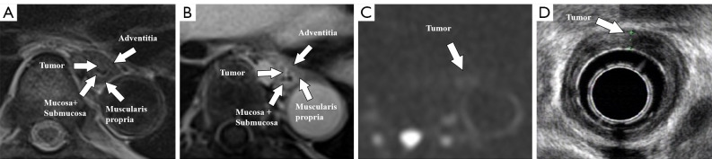
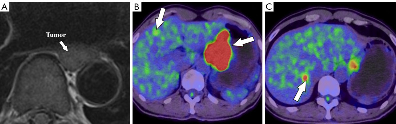
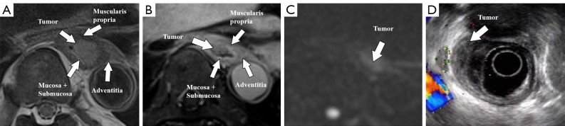
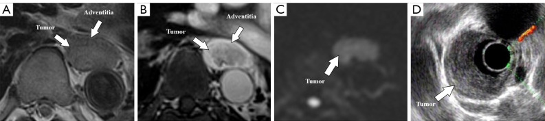
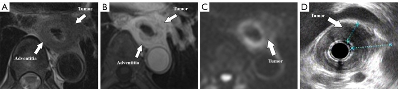
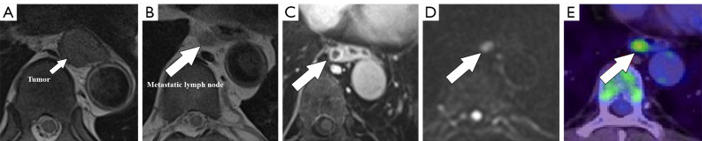
The review highlights the distinct advantages and inherent limitations of different imaging modalities for the preoperative staging of EC. Endoscopic ultrasound (EUS) provides real-time, high-resolution imaging of the esophageal wall but is operator-dependent. Computed tomography (CT) is widely available and non-invasive, but it may lack sensitivity for early T-stage identification. Positron emission tomography (PET)/CT offers accurate assessment of distant metastasis but has limited value in the evaluation of early-stage tumors. With improved techniques, MRI is particularly useful for visualization of tumor infiltration and the surrounding anatomical structures, gaining prominence in preoperative staging of EC.

CONCLUSIONS

Various imaging modalities including EUS, CT, PET/CT, and MRI should be applied as complementary methods for preoperative TNM staging of EC. Notably, high-resolution MRI can overcome motion-related artifacts and provide high-quality images, which may play a more important role in the management of EC in the future.

摘要

背景与目的

食管癌(EC)是一种侵袭性疾病,其特点是死亡率高,且易发生局部区域或远处复发。食管癌的治疗策略和预后评估取决于准确的术前肿瘤-淋巴结-转移(TNM)分期。本综述的目的是阐述各种成像方式在实现食管癌准确术前TNM分期中的作用,特别关注先进的高分辨率磁共振成像(MRI)序列在T分期中的应用,这些序列在增强肿瘤深度和范围的描绘方面已显示出前景。

方法

在PubMed和Web of Science数据库中进行了全面的文献检索。对从这些数据库建立之初至2022年12月31日以英文发表的关于食管癌术前TNM分期成像的研究进行了综述。

关键内容与发现

本综述强调了不同成像方式在食管癌术前分期中的独特优势和固有局限性。内镜超声(EUS)可提供食管壁的实时高分辨率成像,但依赖操作者。计算机断层扫描(CT)广泛可用且无创,但对早期T分期的识别可能缺乏敏感性。正电子发射断层扫描(PET)/CT能准确评估远处转移,但在早期肿瘤评估中的价值有限。随着技术的改进,MRI对肿瘤浸润和周围解剖结构的可视化特别有用,在食管癌术前分期中日益突出。

结论

包括EUS、CT、PET/CT和MRI在内的各种成像方式应作为食管癌术前TNM分期的补充方法应用。值得注意的是,高分辨率MRI可克服与运动相关的伪影并提供高质量图像,未来可能在食管癌的管理中发挥更重要的作用。

https://cdn.ncbi.nlm.nih.gov/pmc/blobs/6c84/11652015/9197bebdf3ff/qims-14-12-9589-f6.jpg
https://cdn.ncbi.nlm.nih.gov/pmc/blobs/6c84/11652015/8b76a15dd2d1/qims-14-12-9589-f1.jpg
https://cdn.ncbi.nlm.nih.gov/pmc/blobs/6c84/11652015/4f54ed5031cb/qims-14-12-9589-f2.jpg
https://cdn.ncbi.nlm.nih.gov/pmc/blobs/6c84/11652015/9460d98140bf/qims-14-12-9589-f3.jpg
https://cdn.ncbi.nlm.nih.gov/pmc/blobs/6c84/11652015/61eed4f94b3b/qims-14-12-9589-f4.jpg
https://cdn.ncbi.nlm.nih.gov/pmc/blobs/6c84/11652015/1b72698ccd7a/qims-14-12-9589-f5.jpg
https://cdn.ncbi.nlm.nih.gov/pmc/blobs/6c84/11652015/9197bebdf3ff/qims-14-12-9589-f6.jpg

相似文献

1
Role of high-resolution magnetic resonance imaging in preoperative tumor-node-metastasis staging evaluation of esophageal cancer: a narrative review.高分辨率磁共振成像在食管癌术前肿瘤-淋巴结-转移分期评估中的作用:一项叙述性综述
Quant Imaging Med Surg. 2024 Dec 5;14(12):9589-9599. doi: 10.21037/qims-24-34. Epub 2024 Oct 18.
2
Ultrasound, CT, MRI, or PET-CT for staging and re-staging of adults with cutaneous melanoma.超声、CT、MRI或PET-CT用于成人皮肤黑色素瘤的分期及再分期。
Cochrane Database Syst Rev. 2019 Jul 1;7(7):CD012806. doi: 10.1002/14651858.CD012806.pub2.
3
A prospective analysis of the diagnostic accuracy of 3 T MRI, CT and endoscopic ultrasound for preoperative T staging of potentially resectable esophageal cancer.前瞻性分析 3T MRI、CT 和内镜超声对潜在可切除食管癌术前 T 分期的诊断准确性。
Cancer Imaging. 2020 Sep 10;20(1):64. doi: 10.1186/s40644-020-00343-w.
4
Clinical implication of PET/MR imaging in preoperative esophageal cancer staging: comparison with PET/CT, endoscopic ultrasonography, and CT.PET/MR 成像在术前食管癌分期中的临床意义:与 PET/CT、内镜超声和 CT 的比较。
J Nucl Med. 2014 Aug;55(8):1242-7. doi: 10.2967/jnumed.114.138974. Epub 2014 May 27.
5
The Role of Magnetic Resonance Imaging and Positron Emission Tomography/Computed Tomography in the Primary Staging of Newly Diagnosed Prostate Cancer: A Systematic Review of the Literature.磁共振成像和正电子发射断层扫描/计算机断层扫描在新诊断前列腺癌的分期中的作用:文献的系统评价。
Eur Urol Oncol. 2021 Jun;4(3):370-395. doi: 10.1016/j.euo.2020.11.002. Epub 2020 Dec 4.
6
Evaluation of preoperative staging for esophageal squamous cell carcinoma.食管鳞状细胞癌术前分期的评估
World J Gastroenterol. 2016 Aug 7;22(29):6683-9. doi: 10.3748/wjg.v22.i29.6683.
7
The Role of Magnetic Resonance Imaging in the Management of Esophageal Cancer.磁共振成像在食管癌管理中的作用。
Cancers (Basel). 2022 Feb 23;14(5):1141. doi: 10.3390/cancers14051141.
8
More advantages in detecting bone and soft tissue metastases from prostate cancer using F-PSMA PET/CT.使用F-PSMA PET/CT检测前列腺癌骨和软组织转移方面有更多优势。
Hell J Nucl Med. 2019 Jan-Apr;22(1):6-9. doi: 10.1967/s002449910952. Epub 2019 Mar 7.
9
Advancing Esophageal Cancer Staging and Restaging: The Role of MRI in Precision Diagnosis.进展期食管癌的分期与再分期:MRI在精准诊断中的作用
Cancers (Basel). 2025 Apr 17;17(8):1351. doi: 10.3390/cancers17081351.
10
The role of ultrasound in primary workup of cervical cancer staging (ESGO, ESTRO, ESP cervical cancer guidelines).超声在宫颈癌分期初步检查中的作用(欧洲妇科肿瘤学会、欧洲放射肿瘤学会、西班牙妇科肿瘤学会宫颈癌指南)
Ceska Gynekol. 2019 Winter;84(1):40-48.

本文引用的文献

1
Global cancer statistics 2022: GLOBOCAN estimates of incidence and mortality worldwide for 36 cancers in 185 countries.2022 年全球癌症统计数据:全球 185 个国家和地区 36 种癌症的发病率和死亡率全球估计数。
CA Cancer J Clin. 2024 May-Jun;74(3):229-263. doi: 10.3322/caac.21834. Epub 2024 Apr 4.
2
Comparison of esophageal cancer survival after neoadjuvant chemoradiotherapy plus surgery versus definitive chemoradiotherapy: A systematic review and meta-analysis.新辅助放化疗加手术与单纯放化疗治疗食管癌的生存比较:系统评价和荟萃分析。
Asian J Surg. 2024 Sep;47(9):3827-3840. doi: 10.1016/j.asjsur.2024.02.099. Epub 2024 Mar 5.
3
Prediction of lymph node metastasis in early esophageal cancer.
早期食管癌淋巴结转移的预测
World J Gastrointest Surg. 2023 Oct 27;15(10):2294-2304. doi: 10.4240/wjgs.v15.i10.2294.
4
F-FDG PET/CT predicts the role of neoadjuvant immunochemotherapy in the pathological response of esophageal squamous cell carcinoma.氟代脱氧葡萄糖正电子发射断层扫描/计算机断层扫描预测新辅助免疫化疗在食管鳞癌病理反应中的作用。
Thorac Cancer. 2023 Aug;14(24):2338-2349. doi: 10.1111/1759-7714.15024. Epub 2023 Jul 9.
5
Esophageal and Esophagogastric Junction Cancers, Version 2.2023, NCCN Clinical Practice Guidelines in Oncology.《食管癌和食管胃交界癌,第2版,2023年,美国国立综合癌症网络肿瘤学临床实践指南》
J Natl Compr Canc Netw. 2023 Apr;21(4):393-422. doi: 10.6004/jnccn.2023.0019.
6
Potential use of high-resolution T2-weighted MRI with histopathologic findings in staging esophageal cancer.高分辨率T2加权磁共振成像结合组织病理学结果在食管癌分期中的潜在应用。
Quant Imaging Med Surg. 2023 Jan 1;13(1):249-258. doi: 10.21037/qims-22-376. Epub 2022 Oct 22.
7
Esophageal Cancer Staging.食管癌分期。
Thorac Surg Clin. 2022 Nov;32(4):437-445. doi: 10.1016/j.thorsurg.2022.06.006.
8
Oesophageal cancer: ESMO Clinical Practice Guideline for diagnosis, treatment and follow-up.食管癌:ESMO 诊断、治疗及随访临床实践指南
Ann Oncol. 2022 Oct;33(10):992-1004. doi: 10.1016/j.annonc.2022.07.003. Epub 2022 Jul 29.
9
The survival of esophageal cancer by subtype in China with comparison to the United States.中国与美国食管癌亚型的生存情况比较。
Int J Cancer. 2023 Jan 15;152(2):151-161. doi: 10.1002/ijc.34232. Epub 2022 Aug 20.
10
Nomogram for prognosis of patients with esophageal squamous cell cancer after minimally invasive esophagectomy established based on non-textbook outcome.基于非传统预后结果建立的微创食管切除术后食管鳞状细胞癌患者预后列线图。
Surg Endosc. 2022 Nov;36(11):8326-8339. doi: 10.1007/s00464-022-09290-y. Epub 2022 May 12.