• 文献检索
  • 文档翻译
  • 深度研究
  • 学术资讯
  • Suppr Zotero 插件Zotero 插件
  • 邀请有礼
  • 套餐&价格
  • 历史记录
应用&插件
Suppr Zotero 插件Zotero 插件浏览器插件Mac 客户端Windows 客户端微信小程序
定价
高级版会员购买积分包购买API积分包
服务
文献检索文档翻译深度研究API 文档MCP 服务
关于我们
关于 Suppr公司介绍联系我们用户协议隐私条款
关注我们

Suppr 超能文献

核心技术专利:CN118964589B侵权必究
粤ICP备2023148730 号-1Suppr @ 2026

文献检索

告别复杂PubMed语法,用中文像聊天一样搜索,搜遍4000万医学文献。AI智能推荐,让科研检索更轻松。

立即免费搜索

文件翻译

保留排版,准确专业,支持PDF/Word/PPT等文件格式,支持 12+语言互译。

免费翻译文档

深度研究

AI帮你快速写综述,25分钟生成高质量综述,智能提取关键信息,辅助科研写作。

立即免费体验

利用三维计算机断层扫描进行软骨发育不全和低软骨发育不全的产前诊断:单机构病例系列研究

Prenatal diagnosis of achondroplasia and hypochondroplasia using three-dimensional computed tomography: a case series at a single institution.

作者信息

Waratani Miyoko, Hasegawa Tatsuji, Shimura Koki, Tanaka Yukiko, Ito Fumitake, Takahata Akiko, Mori Taisuke

机构信息

Department of Obstetrics and Gynecology, Graduate School of Medicine Science, Kyoto Prefectural University of Medicine, Kyoto, Japan.

Department of Pediatrics, Graduate School of Medicine Science, Kyoto Prefectural University of Medicine, Kyoto, Japan.

出版信息

Quant Imaging Med Surg. 2024 Dec 5;14(12):9543-9551. doi: 10.21037/qims-24-682. Epub 2024 Nov 6.

DOI:10.21037/qims-24-682
PMID:39698715
原文链接:https://pmc.ncbi.nlm.nih.gov/articles/PMC11651972/
Abstract

BACKGROUND

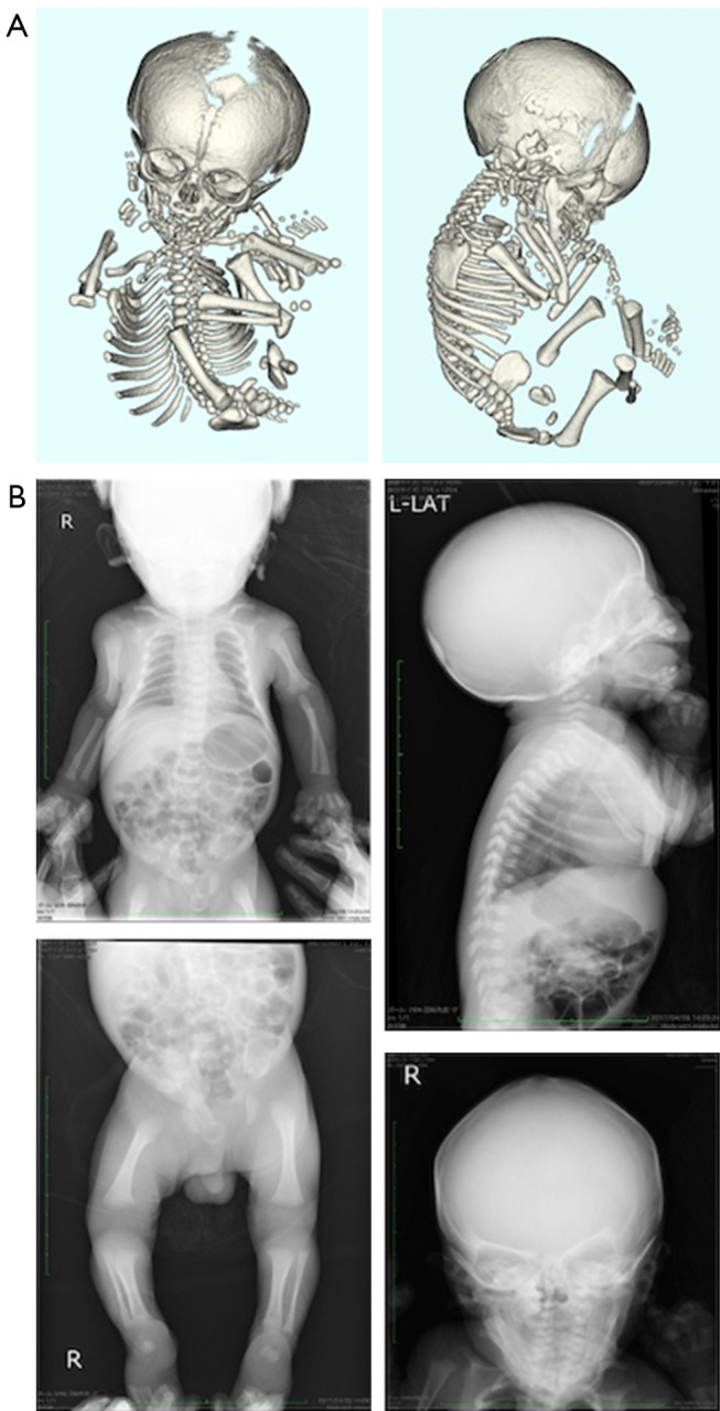
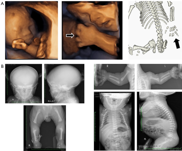
Fetal skeletal dysplasia (FSD) is a group of systemic bone and cartilage disorders that develop prenatally and can be detected using fetal ultrasonography. However, it is unsuitable for skeletal analysis because it is reflected by supersonic waves in the bone cortex. Three-dimensional computed tomography (3D-CT) is a suitable alternative and has improved the differential diagnosis of FSD during pregnancy. Achondroplasia (ACH) and hypochondroplasia (HCH) are the most frequent diseases in the FSD group. This study aimed to determine the efficacy of 3D-CT in the prenatal diagnosis of ACH/HCH.

CASE DESCRIPTION

Patients were selected for the study non-consecutively. Pregnant women who met the following selection criteria were included: (I) pregnancy at age ≥20 years at the time of consent; (II) fetal ultrasonography showing a short femur (below the 10 percentile of fetal measurements) or an image of a long bone curvature or deformity; and (III) written informed consent to undergo 3D-CT. On suspicion of FSD based on ultrasonography, a 3D-CT scan was performed prenatally and an X-ray postnatally to obtain detailed skeletal information and to verify the shapes of all bones, respectively. A genetic examination was performed to confirm the diagnosis after obtaining informed consent from the parents. There were seven cases of prenatally diagnosed ACH/HCH. Genetic testing was performed in six infants postnatally, and a mutation in the fibroblast growth factor receptor 3 () gene was detected [c. 1138G>A (p. gly380Arg)]. In one case, the patient was diagnosed with ACH and Down syndrome by genetic and chromosomal testing (G-band: 47, XY, +21).

CONCLUSIONS

3D-CT is a valuable and efficient tool for diagnosing ACH/HCH. Accurate prenatal diagnosis by gene analysis is crucial for a definitive diagnosis in infants.

摘要

背景

胎儿骨骼发育不良(FSD)是一组产前发生的系统性骨骼和软骨疾病,可通过胎儿超声检查检测到。然而,由于其在骨皮质中由超声波反射显示,所以不适合进行骨骼分析。三维计算机断层扫描(3D-CT)是一种合适的替代方法,已改善了孕期FSD的鉴别诊断。软骨发育不全(ACH)和软骨发育低下(HCH)是FSD组中最常见的疾病。本研究旨在确定3D-CT在ACH/HCH产前诊断中的有效性。

病例描述

非连续选取患者进行本研究。纳入符合以下入选标准的孕妇:(I)同意时年龄≥20岁;(II)胎儿超声检查显示股骨短(低于胎儿测量值的第10百分位数)或长骨弯曲或畸形图像;(III)书面知情同意接受3D-CT检查。基于超声怀疑FSD时,产前进行3D-CT扫描,产后进行X线检查,分别获取详细的骨骼信息并验证所有骨骼的形态。在获得父母知情同意后进行基因检测以确诊。共有7例产前诊断为ACH/HCH的病例。6例婴儿产后进行了基因检测,检测到成纤维细胞生长因子受体3()基因的突变 [c. 1138G>A(p. gly380Arg)]。1例患者通过基因和染色体检测(G带:47,XY,+21)被诊断为ACH和唐氏综合征。

结论

3D-CT是诊断ACH/HCH的一种有价值且有效的工具。通过基因分析进行准确的产前诊断对于婴儿的确切诊断至关重要。

https://cdn.ncbi.nlm.nih.gov/pmc/blobs/c935/11651972/a40279675c74/qims-14-12-9543-f3.jpg
https://cdn.ncbi.nlm.nih.gov/pmc/blobs/c935/11651972/ca9530e90e6e/qims-14-12-9543-f1.jpg
https://cdn.ncbi.nlm.nih.gov/pmc/blobs/c935/11651972/562ef8b59fe2/qims-14-12-9543-f2.jpg
https://cdn.ncbi.nlm.nih.gov/pmc/blobs/c935/11651972/a40279675c74/qims-14-12-9543-f3.jpg
https://cdn.ncbi.nlm.nih.gov/pmc/blobs/c935/11651972/ca9530e90e6e/qims-14-12-9543-f1.jpg
https://cdn.ncbi.nlm.nih.gov/pmc/blobs/c935/11651972/562ef8b59fe2/qims-14-12-9543-f2.jpg
https://cdn.ncbi.nlm.nih.gov/pmc/blobs/c935/11651972/a40279675c74/qims-14-12-9543-f3.jpg

相似文献

1
Prenatal diagnosis of achondroplasia and hypochondroplasia using three-dimensional computed tomography: a case series at a single institution.利用三维计算机断层扫描进行软骨发育不全和低软骨发育不全的产前诊断:单机构病例系列研究
Quant Imaging Med Surg. 2024 Dec 5;14(12):9543-9551. doi: 10.21037/qims-24-682. Epub 2024 Nov 6.
2
Prenatal diagnosis of fetal skeletal dysplasia using 3-dimensional computed tomography: a prospective study.使用三维计算机断层扫描进行胎儿骨骼发育异常的产前诊断:一项前瞻性研究。
BMC Musculoskelet Disord. 2020 Oct 8;21(1):662. doi: 10.1186/s12891-020-03663-x.
3
Detection of a de novo Y278C mutation in FGFR3 in a pregnancy with severe fetal hypochondroplasia: prenatal diagnosis and literature review.一例患有严重胎儿软骨发育不全的妊娠中FGFR3基因新发Y278C突变的检测:产前诊断及文献综述
Taiwan J Obstet Gynecol. 2013 Dec;52(4):580-5. doi: 10.1016/j.tjog.2013.10.023.
4
Low bone mineral density in achondroplasia and hypochondroplasia.软骨发育不全和低软骨发育不全中的低骨矿物质密度。
Pediatr Int. 2016 Aug;58(8):705-8. doi: 10.1111/ped.12890. Epub 2016 Apr 5.
5
p.Ser348Cys mutation in FGFR3 gene leads to "Mild ACH /Severe HCH" phenotype.FGFR3基因中的p.Ser348Cys突变导致“轻度软骨发育不全/重度遗传性软骨发育不全”表型。
Eur J Med Genet. 2020 Feb;63(2):103659. doi: 10.1016/j.ejmg.2019.04.016. Epub 2019 Apr 30.
6
Novel FGFR3 mutations creating cysteine residues in the extracellular domain of the receptor cause achondroplasia or severe forms of hypochondroplasia.在受体细胞外结构域产生半胱氨酸残基的新型FGFR3突变会导致软骨发育不全或严重形式的软骨发育低下。
Eur J Hum Genet. 2006 Dec;14(12):1240-7. doi: 10.1038/sj.ejhg.5201700. Epub 2006 Aug 16.
7
Novel phenotype of achondroplasia due to biallelic FGFR3 pathogenic variants.双等位基因FGFR3致病性变异导致的软骨发育不全新表型。
Am J Med Genet A. 2018 Jul;176(7):1675-1679. doi: 10.1002/ajmg.a.38839.
8
[Clinical analysis of 21 cases with short fetal femur in the third trimester].孕晚期胎儿股骨短小21例临床分析
Zhonghua Fu Chan Ke Za Zhi. 2017 Feb 25;52(2):86-92. doi: 10.3760/cma.j.issn.0529-567X.2017.02.004.
9
FGFR3 mutation frequency in 324 cases from the International Skeletal Dysplasia Registry.国际骨骼发育异常登记处324例病例中的FGFR3突变频率。
Mol Genet Genomic Med. 2014 Nov;2(6):497-503. doi: 10.1002/mgg3.96. Epub 2014 Aug 5.
10
[Clinical analysis and genetic diagnosis of short-limb inherited short stature diseases in children].儿童短肢遗传性矮小症的临床分析与基因诊断
Zhongguo Dang Dai Er Ke Za Zhi. 2013 Nov;15(11):932-6.

本文引用的文献

1
Nosology of genetic skeletal disorders: 2023 revision.遗传骨骼疾病分类学:2023 修订版。
Am J Med Genet A. 2023 May;191(5):1164-1209. doi: 10.1002/ajmg.a.63132. Epub 2023 Feb 13.
2
Achondroplasia: Update on diagnosis, follow-up and treatment.软骨发育不全:诊断、随访及治疗的最新进展
An Pediatr (Engl Ed). 2022 Dec;97(6):423.e1-423.e11. doi: 10.1016/j.anpede.2022.10.004. Epub 2022 Nov 5.
3
Prenatal diagnosis of fetal skeletal dysplasia using 3-dimensional computed tomography: a prospective study.使用三维计算机断层扫描进行胎儿骨骼发育异常的产前诊断:一项前瞻性研究。
BMC Musculoskelet Disord. 2020 Oct 8;21(1):662. doi: 10.1186/s12891-020-03663-x.
4
Novel therapeutic approaches for the treatment of achondroplasia.治疗软骨发育不全症的新疗法。
Bone. 2020 Dec;141:115579. doi: 10.1016/j.bone.2020.115579. Epub 2020 Aug 11.
5
Achondroplasia: Development, pathogenesis, and therapy.软骨发育不全:发育、发病机制及治疗
Dev Dyn. 2017 Apr;246(4):291-309. doi: 10.1002/dvdy.24479. Epub 2017 Mar 2.
6
Skeleton Genetics: a comprehensive database for genes and mutations related to genetic skeletal disorders.骨骼遗传学:一个关于与遗传性骨骼疾病相关的基因和突变的综合数据库。
Database (Oxford). 2016 Aug 31;2016. doi: 10.1093/database/baw127. Print 2016.
7
Use of low dose computed tomography with 3D reconstructions for the prenatal evaluation of suspected skeletal dysplasia.使用低剂量计算机断层扫描及三维重建技术进行疑似骨骼发育异常的产前评估。
Isr Med Assoc J. 2015 Jan;17(1):42-6.
8
Association of achondroplasia with Down syndrome: difficulty in prenatal diagnosis by sonographic and 3-D helical computed tomographic analyses.软骨发育不全与唐氏综合征的关联:超声及三维螺旋计算机断层扫描分析在产前诊断中的困难
Congenit Anom (Kyoto). 2015 May;55(2):116-20. doi: 10.1111/cga.12097.
9
Outcome of fetuses with short femur length detected at second-trimester anomaly scan: a national survey.孕中期异常扫描时检测到股骨长度短的胎儿的结局:一项全国性调查。
Ultrasound Obstet Gynecol. 2014 Aug;44(2):160-5. doi: 10.1002/uog.13286. Epub 2014 Jun 27.
10
Low-dose fetal CT in the prenatal evaluation of skeletal dysplasias and other severe skeletal abnormalities.胎儿 CT 低剂量扫描在骨骼发育不良和其他严重骨骼异常的产前评估中的应用。
AJR Am J Roentgenol. 2013 May;200(5):989-1000. doi: 10.2214/AJR.12.9722.