Alkhatatba Mohammad, Essa Suhaib Bani, Khatatbeh Moawiah, Radaideh Ahmad, Audat Hamzeh Ziad, Younes Ahmad Bani, Alrawashdeh Mutaz, Abualadas Jehad, Obeidat Naser, Al-Omari Jamal, Anaqreh Yazan
Department of Special Surgery, Faculty of Medicine, Jordan University of Science and Technology, Irbid, Jordan.
Department of Public Health, Faculty of Medicine, Yarmouk University Irbid, Jordan.
Mater Sociomed. 2024;36(2):131-136. doi: 10.5455/msm.2024.36.131-136.
Flexible flatfoot is a normal finding in infants and the arch is shaped spontaneously in most children before the age of 10 years. Flexible flatfoot is a common deformity in both adolescent and adult populations.
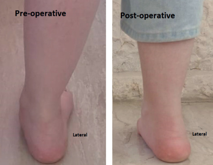
This prospective study aims to assess the functional and radiological outcomes of subtalar arthroereisis in adolescent patients with symptomatic flexible flatfoot.
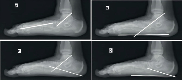
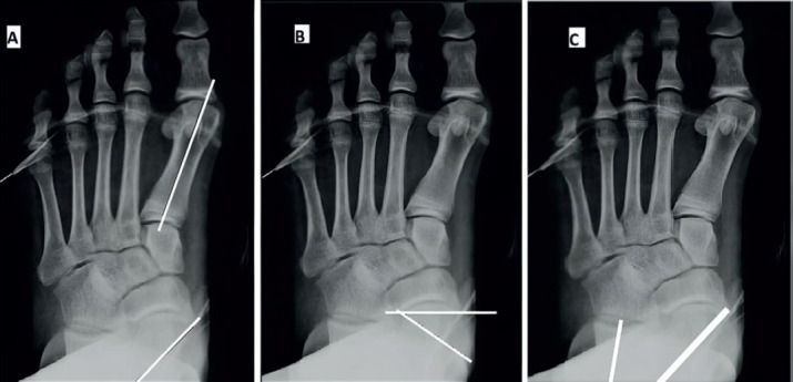
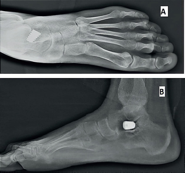
This is a prospective study and included 26 feet in 19 patients who underwent subtalar arthroereisis for symptomatic flexible flatfeet deformity. Preoperative and postoperative functional assessment based on the American Orthopedic Foot and Ankle Society (AOFAS) hindfoot scale. Radiographic parameters included preoperative and postoperative Kites angle, talonavicular coverage angle, Anterior-Posterior talo-1st metatarsal angle, Mearys angle, talar declination angle, calcaneal inclination angle and lateral talocalcaneal angle.
The mean follow-up period was 22.5±9.4 months and the mean preoperative AOFAS score was 54.6±6.0, while the mean AOFAS score at the last follow-up visit was 86.3±3.9 (P<0.001).The mean preoperative and postoperative radiological measurements were 19.0°±8.2° and 7.4°±3.9° for the AP Talo-1st metatarsal angle (P<0.001); 23.6°±9.1° and 8.0°±4.0° for talonavicular coverage angle (P<0.001); 35.4°±3.7° and 24.1°±3.4° for Kites angle (P<0.003); 22.4°±6.1° and 7.5°±3.7° for Mearys angle (P<0.001); 41.0°±4.4° and 25.2°±7.1° for talar declination angle (P<0.001); 13.5°±3.7° and 21.3°±3.6° for calcaneal inclination angle (P<0.001) and 52.4°±7.2° and 42.9°±4.8° for lateral talocalcaneal angle (P<0.041) respectively.
Subtalar arthroereisis is an effective and minimally invasive procedure that showed clinical and radiological improvement for symptomatic flexible flatfoot in our study group.
柔韧性扁平足在婴儿中是正常现象,大多数儿童在10岁前足弓会自然形成。柔韧性扁平足在青少年和成人中都是常见的畸形。
本前瞻性研究旨在评估距下关节制动术治疗有症状的柔韧性扁平足青少年患者的功能和影像学结果。
这是一项前瞻性研究,纳入了19例因有症状的柔韧性扁平足畸形接受距下关节制动术的患者的26只足。基于美国矫形足踝协会(AOFAS)后足评分进行术前和术后功能评估。影像学参数包括术前和术后的Kite角、距舟覆盖角、距骨-第一跖骨前后角、Meary角、距骨倾斜角、跟骨倾斜角和外侧距下关节角。
平均随访期为22.5±9.4个月,术前AOFAS平均评分为54.6±6.0,而末次随访时AOFAS平均评分为86.3±3.9(P<0.001)。距骨-第一跖骨前后角术前和术后的平均影像学测量值分别为19.0°±8.2°和7.4°±3.9°(P<0.001);距舟覆盖角分别为23.6°±9.1°和8.0°±4.0°(P<0.001);Kite角分别为35.4°±3.7°和24.1°±3.4°(P<0.003);Meary角分别为22.4°±6.1°和7.5°±3.7°(P<0.001);距骨倾斜角分别为41.0°±4.4°和25.2°±7.1°(P<0.001);跟骨倾斜角分别为13.5°±3.7°和21.3°±3.6°(P<0.001);外侧距下关节角分别为52.4°±7.2°和42.9°±4.8°(P<0.041)。
距下关节制动术是一种有效且微创的手术,在我们的研究组中,该手术使有症状的柔韧性扁平足在临床和影像学方面均有改善。