Theobald Carina C, Lotfinia Ahmadali, Knobloch Jan A, Medlej Yasser, Stevens David R, Lauterbach Marcel A
Saarland University, Molecular Imaging, Center for Integrative Physiology and Molecular Medicine, Homburg, Germany.
Neurophotonics. 2025 Jan;12(1):015001. doi: 10.1117/1.NPh.12.1.015001. Epub 2024 Dec 19.
Neuronal dendritic spines are central elements for memory and learning. Their morphology correlates with synaptic strength and is a proxy for function. Classic light microscopy cannot resolve spine morphology well, and techniques with higher resolution (electron microscopy and super-resolution light microscopy) typically do not provide spine data in large fields of view, e.g., along entire dendrites. Therefore, it remains unclear if spine types are organized on mesoscopic scales, despite their undisputed importance for understanding the brain.
Recently, it was shown that the distribution of spine type is dendrite-specific in the turtle cortex, suggesting a mesoscopic organization, but leaving the question open if such a dendrite specificity also exists in mammals. Here, we determine if such a difference in spine-type distribution among dendrites also exists in the mouse brain.
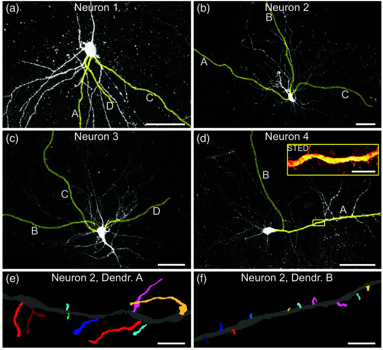
We used super-resolution stimulated emission depletion microscopy of complete dendrites and advanced morphological analysis in three dimensions to decipher morphological differences of spines on different dendrites.
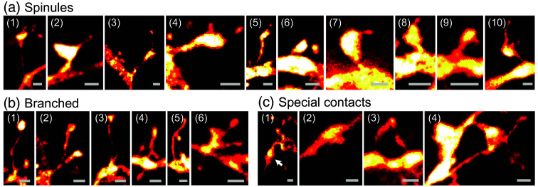
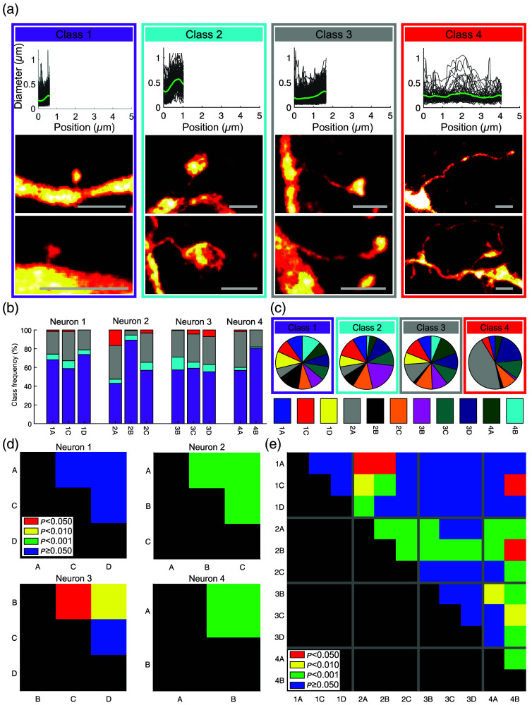
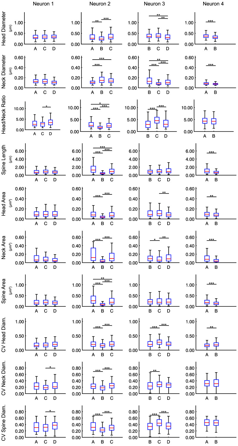
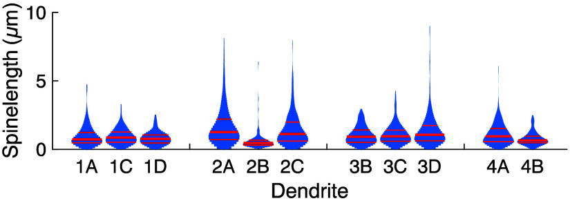
We found that spines of different shapes decorate different dendrites of the same neuron to a varying extent. Significant differences among the dendrites are apparent, based on spine classes as well as based on quantitative descriptors, such as spine length or head size.
Our findings may indicate that it is an evolutionarily conserved principle that individual dendrites have distinct distributions of spine types hinting at individual roles.
神经元树突棘是记忆和学习的核心要素。它们的形态与突触强度相关,是功能的一种指标。传统光学显微镜无法很好地分辨棘的形态,而具有更高分辨率的技术(电子显微镜和超分辨率光学显微镜)通常无法在大视野中提供棘的数据,例如沿整个树突。因此,尽管棘类型对于理解大脑至关重要,但目前仍不清楚它们在介观尺度上是否有组织。
最近有研究表明,在龟脑皮层中棘类型的分布具有树突特异性,这表明存在介观组织,但哺乳动物中是否也存在这种树突特异性仍未可知。在此,我们确定小鼠大脑中不同树突之间是否也存在棘类型分布的差异。
我们使用超分辨率受激发射损耗显微镜观察完整的树突,并进行三维高级形态分析,以解读不同树突上棘的形态差异。
我们发现,不同形状的棘在同一神经元的不同树突上的分布程度各不相同。基于棘的类别以及诸如棘长度或头部大小等定量描述符,不同树突之间存在显著差异。
我们的研究结果可能表明,单个树突具有不同的棘类型分布,暗示着各自的作用,这是一个进化上保守的原则。