• 文献检索
  • 文档翻译
  • 深度研究
  • 学术资讯
  • Suppr Zotero 插件Zotero 插件
  • 邀请有礼
  • 套餐&价格
  • 历史记录
应用&插件
Suppr Zotero 插件Zotero 插件浏览器插件Mac 客户端Windows 客户端微信小程序
定价
高级版会员购买积分包购买API积分包
服务
文献检索文档翻译深度研究API 文档MCP 服务
关于我们
关于 Suppr公司介绍联系我们用户协议隐私条款
关注我们

Suppr 超能文献

核心技术专利:CN118964589B侵权必究
粤ICP备2023148730 号-1Suppr @ 2026

文献检索

告别复杂PubMed语法,用中文像聊天一样搜索,搜遍4000万医学文献。AI智能推荐,让科研检索更轻松。

立即免费搜索

文件翻译

保留排版,准确专业,支持PDF/Word/PPT等文件格式,支持 12+语言互译。

免费翻译文档

深度研究

AI帮你快速写综述,25分钟生成高质量综述,智能提取关键信息,辅助科研写作。

立即免费体验

食管鳞状细胞癌根治性放化疗前后T淋巴细胞亚群上PD-1表达及相关免疫指标的变化

Changes in PD-1 expression on T lymphocyte subsets and related immune indicators before and after definitive chemoradiotherapy for esophageal squamous cell carcinoma.

作者信息

Shi Xueling, Zhao Hongyu, Yu Jiaqi, Cai Peng, Zhou Shixiang, Yang Ning, Li Duojie

机构信息

Department of Radiotherapy, The First Affiliated Hospital of Bengbu Medical University, Bengbu, Anhui, China.

Anhui Province Key Laboratory of Cancer Translational Medicine, Bengbu Medical University, Bengbu, Anhui, China.

出版信息

Ann Med. 2025 Dec;57(1):2445190. doi: 10.1080/07853890.2024.2445190. Epub 2024 Dec 23.

DOI:10.1080/07853890.2024.2445190
PMID:39713872
原文链接:https://pmc.ncbi.nlm.nih.gov/articles/PMC11703528/
Abstract

OBJECTIVE

This study aimed to observe the dynamic changes in the expression of T lymphocytes, natural killer (NK) cells, and PD-1 in patients with first-diagnosed esophageal squamous cell carcinoma (ESCC) before and after chemoradiotherapy (CRT) and evaluate the impact of PD-1 expression in peripheral blood on the short-term outcome of patients with ESCC.

PATIENTS AND METHODS

Seventy-three patients with ESCC who were treated with definitive CRT were enrolled. Before and after CRT, flow cytometry was used to detect thePD-1 expression in the peripheral blood and related immune indicators. Peripheral blood from 10 healthy individuals was used as control.

RESULTS

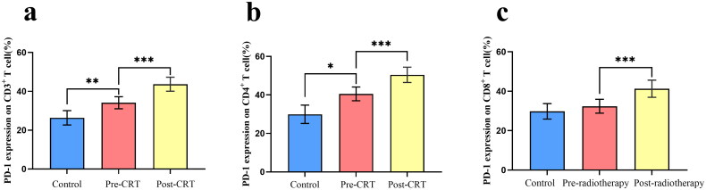
The percentages of CD3 ( = 0.018), CD4 ( < 0.001), and CD8 T cells ( < 0.001); NK cells ( = 0.009); and the CD4/CD8 ratio ( < 0.001), as well as PD-1CD3 ( < 0.001), PD-1CD4 ( < 0.001), and PD-1CD8 ( < 0.001) T cells, before CRT significantly differed from those in the post-CRT group. The percentages of PD-1CD8 T cells differed significantly between the radiotherapy alone and CRT groups ( < 0.05). PD-1 expression in CD3, CD4, and CD8 T cells significantly decreased in patients achieving overall response rate (all  < 0.05). Compared with those in the incomplete response group, PD-1CD8 T cells significantly decreased in the CR group ( < 0.05).

CONCLUSION

CRT aggravated immunosuppression and increased PD-1 expression in T lymphocyte subsets in patients with ESCC, possibly related to the radiation field. PD-1 expression in T lymphocyte subsets can predict short-term outcomes in patients and provide a theoretical basis for the sequential application of PD-1 immunosuppressants after radiotherapy and chemotherapy.

摘要

目的

本研究旨在观察初诊食管鳞状细胞癌(ESCC)患者放化疗(CRT)前后T淋巴细胞、自然杀伤(NK)细胞及PD-1表达的动态变化,并评估外周血中PD-1表达对ESCC患者短期预后的影响。

患者与方法

纳入73例行根治性CRT治疗的ESCC患者。CRT前后,采用流式细胞术检测外周血中PD-1表达及相关免疫指标。选取10名健康个体的外周血作为对照。

结果

CRT前,CD3(=0.018)、CD4(<0.001)和CD8 T细胞(<0.001)、NK细胞(=0.009)的百分比,以及CD4/CD8比值(<0.001),还有PD-1⁺CD3(<0.001)、PD-1⁺CD4(<0.001)和PD-1⁺CD8(<0.001)T细胞与CRT后组相比有显著差异。单纯放疗组与CRT组之间PD-1⁺CD8 T细胞百分比有显著差异(<0.05)。达到总体缓解率的患者中,CD3、CD4和CD8 T细胞中的PD-1表达显著降低(均<0.05)。与部分缓解组相比,完全缓解(CR)组中PD-1⁺CD8 T细胞显著减少(<0.05)。

结论

CRT加重了ESCC患者的免疫抑制并增加了T淋巴细胞亚群中PD-1的表达,可能与放射野有关。T淋巴细胞亚群中的PD-1表达可预测患者的短期预后,并为放化疗后序贯应用PD-1免疫抑制剂提供理论依据。

https://cdn.ncbi.nlm.nih.gov/pmc/blobs/32bd/11703528/99fc26c79786/IANN_A_2445190_F0004_B.jpg
https://cdn.ncbi.nlm.nih.gov/pmc/blobs/32bd/11703528/ea0aad3f58a2/IANN_A_2445190_F0001_C.jpg
https://cdn.ncbi.nlm.nih.gov/pmc/blobs/32bd/11703528/d1ade4d20250/IANN_A_2445190_F0002_C.jpg
https://cdn.ncbi.nlm.nih.gov/pmc/blobs/32bd/11703528/5f4901b4e844/IANN_A_2445190_F0003_C.jpg
https://cdn.ncbi.nlm.nih.gov/pmc/blobs/32bd/11703528/99fc26c79786/IANN_A_2445190_F0004_B.jpg

相似文献

1
Changes in PD-1 expression on T lymphocyte subsets and related immune indicators before and after definitive chemoradiotherapy for esophageal squamous cell carcinoma.食管鳞状细胞癌根治性放化疗前后T淋巴细胞亚群上PD-1表达及相关免疫指标的变化
Ann Med. 2025 Dec;57(1):2445190. doi: 10.1080/07853890.2024.2445190. Epub 2024 Dec 23.
2
Definitive chemoradiotherapy induces T-cell-inflamed tumor microenvironment in unresectable locally advanced esophageal squamous cell carcinoma.根治性放化疗可诱导不可切除局部晚期食管鳞癌的 T 细胞炎症肿瘤微环境。
J Gastroenterol. 2024 Sep;59(9):798-811. doi: 10.1007/s00535-024-02120-z. Epub 2024 May 31.
3
Effects of chemoradiotherapy on surface PD-L1 expression in esophageal cancer and its implications for immunotherapy.放化疗对食管癌表面程序性死亡受体配体1(PD-L1)表达的影响及其对免疫治疗的意义
Front Immunol. 2024 Dec 23;15:1509051. doi: 10.3389/fimmu.2024.1509051. eCollection 2024.
4
Changes in Indoleamine 2,3-Dioxygenase 1 Expression and CD8+ Tumor-Infiltrating Lymphocytes after Neoadjuvant Chemoradiation Therapy and Prognostic Significance in Esophageal Squamous Cell Carcinoma.新辅助放化疗后吲哚胺 2,3-双加氧酶 1 表达和 CD8+肿瘤浸润淋巴细胞的变化及其对食管鳞癌的预后意义。
Int J Radiat Oncol Biol Phys. 2020 Sep 1;108(1):286-294. doi: 10.1016/j.ijrobp.2020.01.020. Epub 2020 Jan 28.
5
TENERGY: multicenter phase II study of Atezolizumab monotherapy following definitive Chemoradiotherapy with 5-FU plus Cisplatin in patients with unresectable locally advanced esophageal squamous cell carcinoma.TENERGY:特瑞普利单抗单药治疗联合 5-FU 顺铂放化疗用于不可切除局部晚期食管鳞癌的多中心 II 期研究。
BMC Cancer. 2020 Apr 20;20(1):336. doi: 10.1186/s12885-020-06716-5.
6
Prognostic value of immuno-inflammatory biomarkers in esophageal squamous cell carcinoma patients receiving immunotherapy combined with chemoradiotherapy and its association with immuno-genomic landscape.免疫炎症生物标志物在接受免疫治疗联合放化疗的食管鳞状细胞癌患者中的预后价值及其与免疫基因组格局的关联
BMC Cancer. 2024 Dec 18;24(1):1518. doi: 10.1186/s12885-024-13298-z.
7
PD-L1 expression, CD8+ and CD4+ lymphocyte rate are predictive of pathological complete response after neoadjuvant chemoradiotherapy for squamous cell cancer of the thoracic esophagus.PD-L1 表达、CD8+和 CD4+淋巴细胞比率与胸段食管鳞癌新辅助放化疗后病理完全缓解相关。
Cancer Med. 2019 Oct;8(13):6036-6048. doi: 10.1002/cam4.2359. Epub 2019 Aug 20.
8
Radiotherapy plus camrelizumab affects peripheral CD8 T-cell differentiation subsets expressing PD-1, TIGIT, and CTLA-4 in esophageal squamous cell carcinoma.放疗联合卡瑞利珠单抗影响食管鳞状细胞癌中表达程序性死亡受体1(PD-1)、T细胞免疫球蛋白和ITIM结构域(TIGIT)以及细胞毒性T淋巴细胞相关蛋白4(CTLA-4)的外周CD8 T细胞分化亚群。
J Leukoc Biol. 2023 Jan 10;113(1):11-17. doi: 10.1093/jleuko/qiac018.
9
Clinical utility of the systemic immune-inflammation index for predicting survival in esophageal squamous cell carcinoma after radical radiotherapy.系统免疫炎症指数对根治性放疗后食管鳞癌患者生存的预测价值。
Future Oncol. 2021 Jul;17(20):2647-2657. doi: 10.2217/fon-2021-0304. Epub 2021 May 19.
10
Dual TGF-β and PD-1 blockade synergistically enhances MAGE-A3-specific CD8 T cell response in esophageal squamous cell carcinoma.双重 TGF-β 和 PD-1 阻断协同增强食管鳞癌中 MAGE-A3 特异性 CD8 T 细胞反应。
Int J Cancer. 2018 Nov 15;143(10):2561-2574. doi: 10.1002/ijc.31730. Epub 2018 Sep 21.

本文引用的文献

1
Modulation of CD8 T Cell Responses by Radiotherapy-Current Evidence and Rationale for Combination with Immune Checkpoint Inhibitors.放疗调控 CD8 T 细胞反应:联合免疫检查点抑制剂的当前证据和原理。
Int J Mol Sci. 2023 Nov 24;24(23):16691. doi: 10.3390/ijms242316691.
2
Mechanisms of radiotherapy resistance and radiosensitization strategies for esophageal squamous cell carcinoma.放射治疗抵抗的机制及食管鳞癌的放射增敏策略。
Mol Cancer. 2023 Aug 19;22(1):140. doi: 10.1186/s12943-023-01839-2.
3
Changes in T lymphocyte subsets in peripheral blood of patients with middle-advanced cervical cancer before and after nimotuzumab combined with concurrent chemoradiotherapy.
尼妥珠单抗联合放化疗治疗中晚期宫颈癌患者前后外周血 T 淋巴细胞亚群的变化。
J Obstet Gynaecol. 2023 Dec;43(1):2179915. doi: 10.1080/01443615.2023.2179915.
4
Radiotherapy plus camrelizumab affects peripheral CD8 T-cell differentiation subsets expressing PD-1, TIGIT, and CTLA-4 in esophageal squamous cell carcinoma.放疗联合卡瑞利珠单抗影响食管鳞状细胞癌中表达程序性死亡受体1(PD-1)、T细胞免疫球蛋白和ITIM结构域(TIGIT)以及细胞毒性T淋巴细胞相关蛋白4(CTLA-4)的外周CD8 T细胞分化亚群。
J Leukoc Biol. 2023 Jan 10;113(1):11-17. doi: 10.1093/jleuko/qiac018.
5
Short-term prognostic role of peripheral lymphocyte subsets in patients with gastric cancer.外周血淋巴细胞亚群在胃癌患者中的短期预后作用
Int Immunopharmacol. 2023 Feb;115:109641. doi: 10.1016/j.intimp.2022.109641. Epub 2022 Dec 28.
6
Comparison of dynamic changes in the peripheral CD8 T cells function and differentiation in ESCC patients treated with radiotherapy combined with anti-PD-1 antibody or concurrent chemoradiotherapy.比较放疗联合抗 PD-1 抗体或同期放化疗治疗的 ESCC 患者外周血 CD8+T 细胞功能和分化的动态变化。
Front Immunol. 2022 Nov 21;13:1060695. doi: 10.3389/fimmu.2022.1060695. eCollection 2022.
7
Peripheral blood lymphocyte subsets are associated with the clinical outcomes of prostate cancer patients.外周血淋巴细胞亚群与前列腺癌患者的临床结局相关。
Int Immunopharmacol. 2022 Dec;113(Pt A):109287. doi: 10.1016/j.intimp.2022.109287. Epub 2022 Oct 14.
8
PD-1/PD-L1 immune-checkpoint blockade induces immune effector cell modulation in metastatic non-small cell lung cancer patients: A single-cell flow cytometry approach.PD-1/PD-L1免疫检查点阻断在转移性非小细胞肺癌患者中诱导免疫效应细胞调节:一种单细胞流式细胞术方法。
Front Oncol. 2022 Sep 14;12:911579. doi: 10.3389/fonc.2022.911579. eCollection 2022.
9
The tissue-resident marker CD103 on peripheral blood T cells predicts responses to anti-PD-1 therapy in gastric cancer.外周血 T 细胞上的组织驻留标志物 CD103 可预测胃癌患者对抗 PD-1 治疗的反应。
Cancer Immunol Immunother. 2023 Jan;72(1):169-181. doi: 10.1007/s00262-022-03240-2. Epub 2022 Jul 1.
10
Mechanistic convergence of the TIGIT and PD-1 inhibitory pathways necessitates co-blockade to optimize anti-tumor CD8 T cell responses.TIGIT 和 PD-1 抑制途径的机制趋同需要联合阻断以优化抗肿瘤 CD8 T 细胞反应。
Immunity. 2022 Mar 8;55(3):512-526.e9. doi: 10.1016/j.immuni.2022.02.005.