Department of Radiation Oncology, Tianjin Medical University Cancer Institute and Hospital, National Clinical Research Center for Cancer, Key Laboratory of Cancer Prevention and Therapy, Tianjin's Clinical Research Center for Cancer, Tianjin, China.
Department of Immunology, Tianjin Medical University Cancer Institute and Hospital, National Clinical Research Center for Cancer, Key Laboratory of Cancer Prevention and Therapy, Tianjin's Clinical Research Center for Cancer, Tianjin, China.
Front Immunol. 2022 Nov 21;13:1060695. doi: 10.3389/fimmu.2022.1060695. eCollection 2022.
The systematic immune status of cancer patients undergoing immunotherapy is little known. We prospectively identified the function and differentiation traits of peripheral CD8 T cells based on our phase 1b clinical trial (NCT03222440) of radiotherapy combined with camrelizumab in patients with locally advanced esophageal squamous cell carcinoma (ESCC) and compared it with concurrent chemoradiotherapy (CCRT).
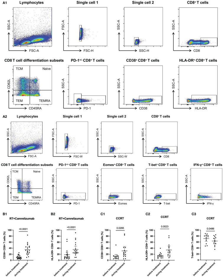
19 and 18 patients were included in the cohort of radiotherapy plus camrelizumab and cohort of CCRT treatment. By using flow cytometry, we evaluated the expression levels of PD-1, Eomes, T-bet and IFN-γ (function), CD38 and HLA-DR (activation), and differentiation subsets classified according to the expression levels of CD45RA and CD62L in peripheral CD8 T cells before and during treatment.
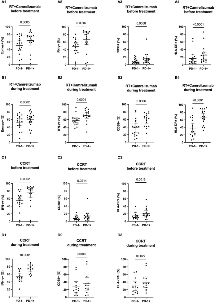
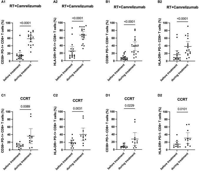
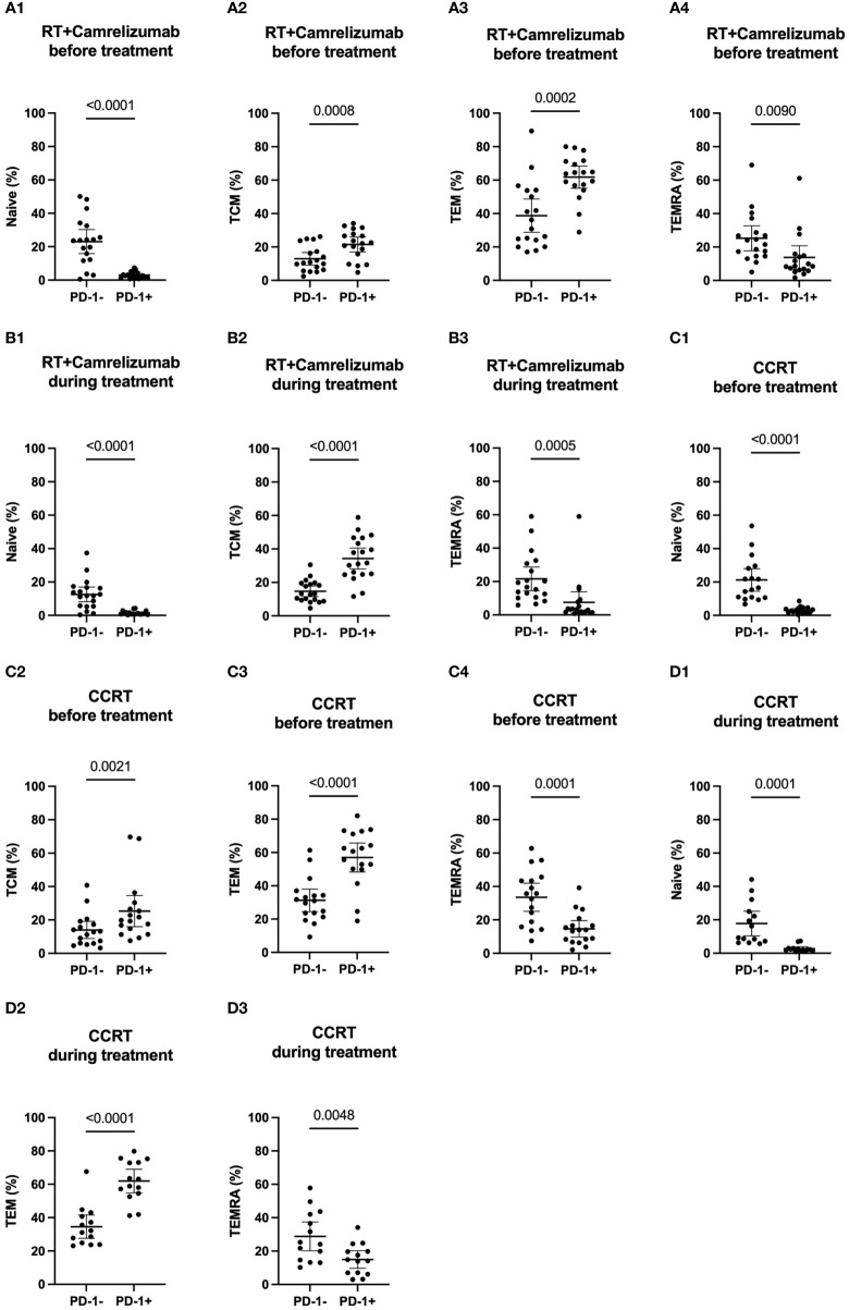
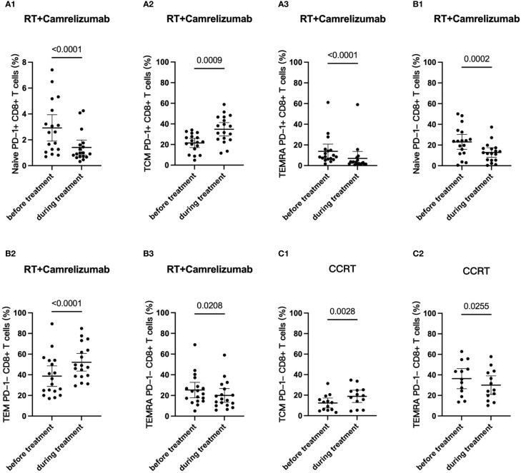
Effective binding of anti-PD-1 antibody camrelizumab with PD-1 on CD8 T cells was detected during treatment. Both two treatments elevated the expression levels of activation molecules CD38 and HLA-DR on CD8 T cells. PD-1CD8 T cells had more activation features than PD-1CD8 T cells in two groups and the treatments did not alter these differences. The two treatments activated both PD-1 and PD-1 CD8 T cells. PD-1CD8 T cells had less Naïve and TEMRA but more Tcm and Tem than PD-1CD8 T cells in two groups and both two treatments changed the ratio of memory T cells in PD-1 and PD-1 cells. RT plus camrelizumab treatment reduced Naïve T cells and TEMRA subsets both in PD-1 and PD-1 CD8 T cells while elevated Tcm subset in PD-1CD8 T cells and Tem subset in PD-1CD8 T cells. CCRT elevated Tcm subset and reduced TEMRA subset in PD-1CD8 T cells while did not change any subset in PD-1CD8 T cells. Furthermore, patients undergoing radiotherapy plus immunotherapy were found to obtain better prognosis than those receiving CCRT.
This study identified the dynamic changes of systematic immune status of patients undergoing treatment. The two treatments had similar activation effects on peripheral CD8 T cells with different PD-1 properties but had different effects on their differentiation status. These results provided potential clues to the reasons underlying the difference in prognosis of the two treatments.
癌症患者接受免疫治疗时的系统性免疫状态知之甚少。我们前瞻性地根据我们的 1b 期临床试验(NCT03222440)确定了接受放射治疗联合卡瑞利珠单抗的局部晚期食管鳞癌(ESCC)患者外周血 CD8 T 细胞的功能和分化特征,并将其与同期放化疗(CCRT)进行了比较。
19 例和 18 例患者分别纳入放疗加卡瑞利珠单抗组和 CCRT 治疗组。通过流式细胞术,我们评估了治疗前后外周血 CD8 T 细胞中 PD-1、Eomes、T-bet 和 IFN-γ(功能)、CD38 和 HLA-DR(激活)以及根据 CD45RA 和 CD62L 表达水平分类的分化亚群的表达水平。
在治疗过程中检测到抗 PD-1 抗体卡瑞利珠单抗与 CD8 T 细胞上 PD-1 的有效结合。两种治疗方法均提高了 CD8 T 细胞上激活分子 CD38 和 HLA-DR 的表达水平。两组中 PD-1+CD8 T 细胞具有更多的激活特征,且两种治疗均未改变这些差异。两种治疗均激活了 PD-1+和 PD-1-CD8 T 细胞。两组中 PD-1+CD8 T 细胞的 Naive 和 TEMRA 亚群较少,而 Tcm 和 Tem 亚群较多,两种治疗均改变了 PD-1 和 PD-1 细胞中记忆 T 细胞的比例。RT 加卡瑞利珠单抗治疗降低了 PD-1 和 PD-1 CD8 T 细胞中 Naive T 细胞和 TEMRA 亚群,而升高了 PD-1 CD8 T 细胞中的 Tcm 亚群和 PD-1 CD8 T 细胞中的 Tem 亚群。CCRT 升高了 PD-1+CD8 T 细胞中的 Tcm 亚群并降低了 PD-1+CD8 T 细胞中的 TEMRA 亚群,但对 PD-1 CD8 T 细胞中的任何亚群均无影响。此外,与接受 CCRT 的患者相比,接受放疗联合免疫治疗的患者预后更好。
本研究确定了接受治疗的患者系统性免疫状态的动态变化。两种治疗方法对具有不同 PD-1 特性的外周血 CD8 T 细胞具有相似的激活作用,但对其分化状态的影响不同。这些结果为两种治疗方法预后差异的原因提供了潜在线索。