• 文献检索
  • 文档翻译
  • 深度研究
  • 学术资讯
  • Suppr Zotero 插件Zotero 插件
  • 邀请有礼
  • 套餐&价格
  • 历史记录
应用&插件
Suppr Zotero 插件Zotero 插件浏览器插件Mac 客户端Windows 客户端微信小程序
定价
高级版会员购买积分包购买API积分包
服务
文献检索文档翻译深度研究API 文档MCP 服务
关于我们
关于 Suppr公司介绍联系我们用户协议隐私条款
关注我们

Suppr 超能文献

核心技术专利:CN118964589B侵权必究
粤ICP备2023148730 号-1Suppr @ 2026

文献检索

告别复杂PubMed语法,用中文像聊天一样搜索,搜遍4000万医学文献。AI智能推荐,让科研检索更轻松。

立即免费搜索

文件翻译

保留排版,准确专业,支持PDF/Word/PPT等文件格式,支持 12+语言互译。

免费翻译文档

深度研究

AI帮你快速写综述,25分钟生成高质量综述,智能提取关键信息,辅助科研写作。

立即免费体验

利用T2WI和DWI MR序列的瘤内及瘤周影像组学特征对乳腺癌组织学分级进行术前预测

Preoperative Prediction of Breast Cancer Histological Grade Using Intratumoral and Peritumoral Radiomics Features from T2WI and DWI MR Sequences.

作者信息

Guo Yaxin, Liao Jun, Li Shunian, Shang Yiyan, Wang Yunxia, Wu Qingxia, Wu Yaping, Wang Meiyun, Yan Fengshan, Tan Hongna

机构信息

Department of Radiology, People's Hospital of Zhengzhou University & Henan Provincial People's Hospital, Zhengzhou, People's Republic of China.

Department of Radiology, People's Hospital of Henan University, Zhengzhou, People's Republic of China.

出版信息

Breast Cancer (Dove Med Press). 2024 Dec 19;16:981-991. doi: 10.2147/BCTT.S487988. eCollection 2024.

DOI:10.2147/BCTT.S487988
PMID:39720357
原文链接:https://pmc.ncbi.nlm.nih.gov/articles/PMC11668253/
Abstract

BACKGROUND

Histological grade is an acknowledged prognostic factor for breast cancer, essential for determining clinical treatment strategies and prognosis assessment. Our study aims to establish intra- and peritumoral radiomics models using T2WI and DWI MR sequences for predicting the histological grade of breast cancer.

METHODS

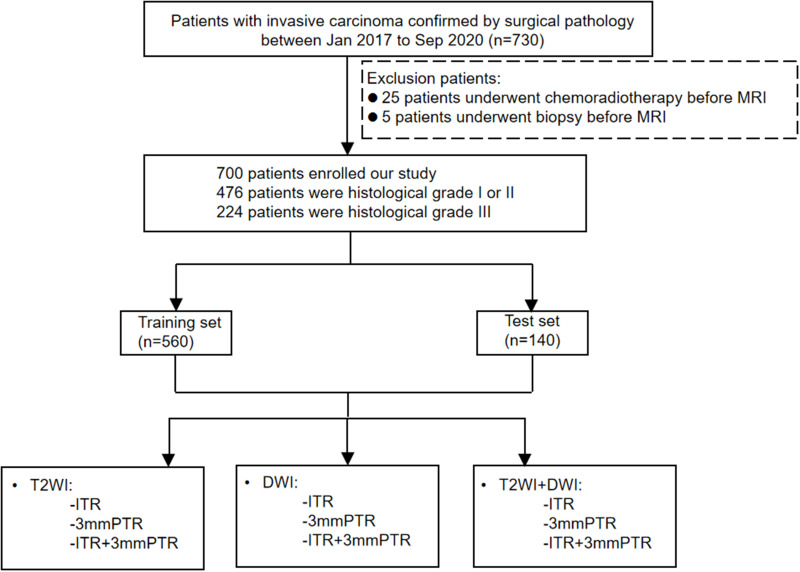
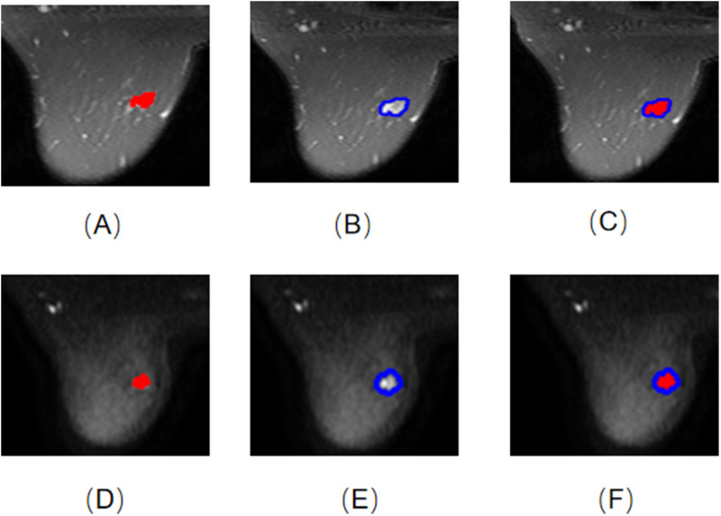
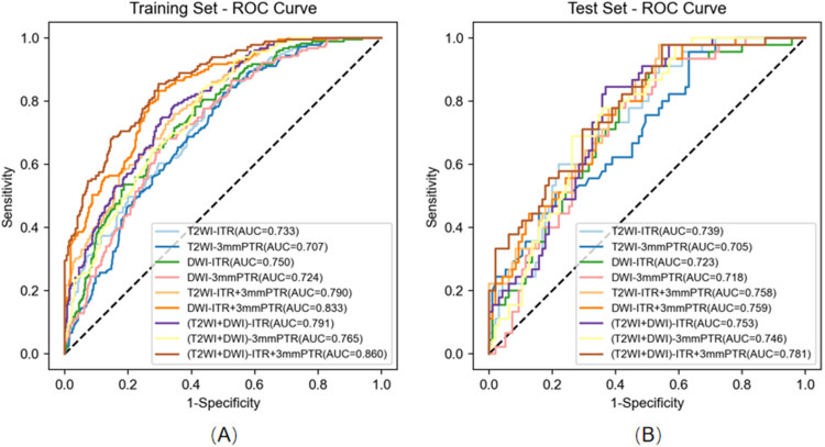
700 breast cancer cases who had MRI scans before surgery were included. The intratumoral region (ITR) of interest was manually delineated, while the peritumoral region (PTR-3 mm) was automatically obtained by expanding the ITR by 3 mm. Radiomics features were extracted using the intra- and peritumoral images from T2WI and DWI sequences on breast MRI. Then, the key features with the strongest predictivity of histological grade were selected. Finally, 9 predictive radiomics models were established based on T2WI-ITR, T2WI-3mmPTR, DWI-ITR, DWI-3mmPTR, T2WI-ITR + 3mmPTR, DWI-ITR + 3mmPTR, (T2WI + DWI)-ITR, (T2WI + DWI)-3mmPTR and (T2WI + DWI)-ITR + 3mmPTR.

RESULTS

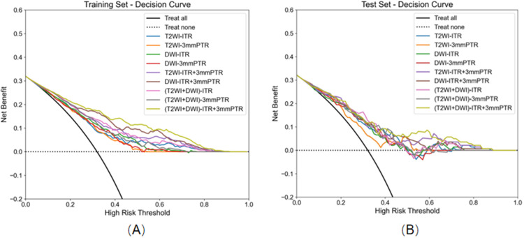
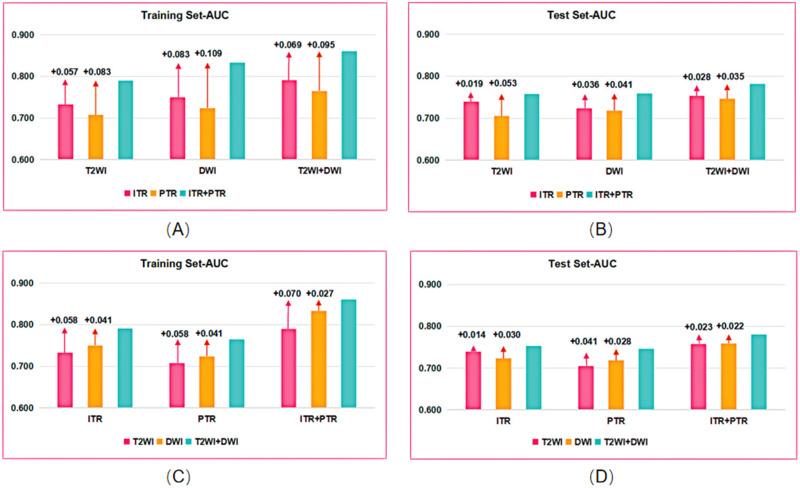
The (T2WI + DWI)-ITR + 3mmPTR contained 13 DWI features which included a shape feature, a texture feature, and 11 filtered features, as well as 10 T2WI features, all of which were filtered features. Among the 9 models, the combined models showed better performance than the single models in both the training and test sets, especially for the (T2WI + DWI)-ITR + 3mmPTR radiomics model. The (T2WI + DWI)-ITR + 3mmPTR radiomics model achieved a sensitivity, specificity, accuracy, and AUC of 80.4%, 72.4%, 75.0%, and 0.860 in the training set, and 68.9%, 70.5%, 70.0%, and 0.781 in the test set. Decision curve analysis (DCA) showed that the (T2WI + DWI)-ITR + 3mmPTR model had the greatest net clinical benefit compared to the other models.

CONCLUSION

The intra- and peritumoral radiomics methodologies using T2WI and DWI MR sequences could be utilized to assess histological grade for breast cancer, particularly with the (T2WI + DWI)-ITR + 3mmPTR radiomics model demonstrating significant potential for clinical application.

摘要

背景

组织学分级是公认的乳腺癌预后因素,对确定临床治疗策略和预后评估至关重要。我们的研究旨在利用T2WI和DWI MR序列建立瘤内和瘤周放射组学模型,以预测乳腺癌的组织学分级。

方法

纳入700例术前进行过MRI扫描的乳腺癌病例。手动勾勒出感兴趣的瘤内区域(ITR),而瘤周区域(PTR-3mm)通过将ITR扩大3mm自动获得。使用乳腺MRI上T2WI和DWI序列的瘤内和瘤周图像提取放射组学特征。然后,选择对组织学分级预测性最强的关键特征。最后,基于T2WI-ITR、T2WI-3mmPTR、DWI-ITR、DWI-3mmPTR、T2WI-ITR + 3mmPTR、DWI-ITR + 3mmPTR、(T2WI + DWI)-ITR、(T2WI + DWI)-3mmPTR和(T2WI + DWI)-ITR + 3mmPTR建立了9个预测性放射组学模型。

结果

(T2WI + DWI)-ITR + 3mmPTR包含13个DWI特征,其中包括1个形状特征、1个纹理特征和11个过滤特征,以及10个T2WI特征,均为过滤特征。在9个模型中,联合模型在训练集和测试集中均表现出比单一模型更好的性能,特别是(T2WI + DWI)-ITR + 3mmPTR放射组学模型。(T2WI + DWI)-ITR + 3mmPTR放射组学模型在训练集中的灵敏度、特异度、准确度和AUC分别为80.4%、72.4%、75.0%和0.860,在测试集中分别为68.9%、70.5%、70.0%和0.781。决策曲线分析(DCA)表明,与其他模型相比,(T2WI + DWI)-ITR + 3mmPTR模型具有最大的净临床效益。

结论

利用T2WI和DWI MR序列的瘤内和瘤周放射组学方法可用于评估乳腺癌的组织学分级,特别是(T2WI + DWI)-ITR + 3mmPTR放射组学模型具有显著的临床应用潜力。

https://cdn.ncbi.nlm.nih.gov/pmc/blobs/1270/11668253/09222f190845/BCTT-16-981-g0005.jpg
https://cdn.ncbi.nlm.nih.gov/pmc/blobs/1270/11668253/59764ff29557/BCTT-16-981-g0001.jpg
https://cdn.ncbi.nlm.nih.gov/pmc/blobs/1270/11668253/7b04ed524e88/BCTT-16-981-g0002.jpg
https://cdn.ncbi.nlm.nih.gov/pmc/blobs/1270/11668253/5a3707d72315/BCTT-16-981-g0003.jpg
https://cdn.ncbi.nlm.nih.gov/pmc/blobs/1270/11668253/78a1a1c3d7fa/BCTT-16-981-g0004.jpg
https://cdn.ncbi.nlm.nih.gov/pmc/blobs/1270/11668253/09222f190845/BCTT-16-981-g0005.jpg

相似文献

1
Preoperative Prediction of Breast Cancer Histological Grade Using Intratumoral and Peritumoral Radiomics Features from T2WI and DWI MR Sequences.利用T2WI和DWI MR序列的瘤内及瘤周影像组学特征对乳腺癌组织学分级进行术前预测
Breast Cancer (Dove Med Press). 2024 Dec 19;16:981-991. doi: 10.2147/BCTT.S487988. eCollection 2024.
2
Clinical-radiomics nomogram based on the fat-suppressed T2 sequence for differentiating luminal and non-luminal breast cancer.基于脂肪抑制T2序列的临床影像组学列线图用于鉴别管腔型和非管腔型乳腺癌。
Front Oncol. 2024 Oct 25;14:1451414. doi: 10.3389/fonc.2024.1451414. eCollection 2024.
3
Clinical study on the prediction of ALN metastasis based on intratumoral and peritumoral DCE-MRI radiomics and clinico-radiological characteristics in breast cancer.基于肿瘤内及瘤周动态对比增强磁共振成像(DCE-MRI)影像组学和临床放射学特征预测乳腺癌腋窝淋巴结转移的临床研究
Front Oncol. 2024 Mar 19;14:1357145. doi: 10.3389/fonc.2024.1357145. eCollection 2024.
4
An Integrative Clinical and Intra- and Peritumoral MRI Radiomics Nomogram for the Preoperative Prediction of Lymphovascular Invasion in Rectal Cancer.一种用于术前预测直肠癌淋巴管侵犯的综合临床及瘤内和瘤周MRI影像组学列线图
Acad Radiol. 2025 Mar 4. doi: 10.1016/j.acra.2025.02.019.
5
Intra- and peri-tumoral MRI radiomics features for preoperative lymph node metastasis prediction in early-stage cervical cancer.用于早期宫颈癌术前淋巴结转移预测的瘤内及瘤周MRI影像组学特征
Insights Imaging. 2023 Apr 15;14(1):65. doi: 10.1186/s13244-023-01405-w.
6
Prediction of bone metastasis of prostate cancer based on intratumoral and peritumoral radiomics of MRI T2WI combined with ADC images.基于MRI T2WI联合ADC图像的肿瘤内及肿瘤周围影像组学预测前列腺癌骨转移
Front Oncol. 2025 Mar 10;15:1555315. doi: 10.3389/fonc.2025.1555315. eCollection 2025.
7
Dual-region MRI radiomic analysis indicates increased risk in high-risk breast lesions: bridging intratumoral and peritumoral radiomics for precision decision-making.双区域MRI影像组学分析表明高危乳腺病变风险增加:连接瘤内和瘤周影像组学以实现精准决策
BMC Cancer. 2025 May 6;25(1):828. doi: 10.1186/s12885-025-14165-1.
8
[Application of MRI-based Radiomics Models in the Assessment of Hepatic Metastasis of Rectal Cancer].基于MRI的影像组学模型在直肠癌肝转移评估中的应用
Sichuan Da Xue Xue Bao Yi Xue Ban. 2021 Mar;52(2):311-318. doi: 10.12182/20210360202.
9
MRI-based multiregional radiomics for preoperative prediction of tumor deposit and prognosis in resectable rectal cancer: a bicenter study.基于 MRI 的多区域放射组学术前预测可切除直肠癌肿瘤沉积和预后的研究:一项双中心研究。
Eur Radiol. 2023 Nov;33(11):7561-7572. doi: 10.1007/s00330-023-09723-9. Epub 2023 May 9.
10
The value of intratumoral and peritumoral radiomics features based on multiparametric MRI for predicting molecular staging of breast cancer.基于多参数磁共振成像的瘤内及瘤周影像组学特征对乳腺癌分子分期的预测价值。
Front Oncol. 2025 Mar 11;15:1379048. doi: 10.3389/fonc.2025.1379048. eCollection 2025.

引用本文的文献

1
Establishment of a predictive nomogram for breast cancer lympho-vascular invasion based on radiomics obtained from digital breast tomography and clinical imaging features.基于数字乳腺断层摄影获得的影像组学和临床影像特征建立乳腺癌淋巴管侵犯的预测列线图。
BMC Med Imaging. 2025 Feb 26;25(1):65. doi: 10.1186/s12880-025-01607-2.

本文引用的文献

1
Optimizing breast cancer diagnosis with photoacoustic imaging: An analysis of intratumoral and peritumoral radiomics.利用光声成像优化乳腺癌诊断:瘤内和瘤周放射组学分析
Photoacoustics. 2024 Apr 9;38:100606. doi: 10.1016/j.pacs.2024.100606. eCollection 2024 Aug.
2
Ultrasound-based radiomics nomogram for predicting axillary lymph node metastasis in early-stage breast cancer.基于超声的放射组学列线图预测早期乳腺癌腋窝淋巴结转移
Radiol Med. 2024 Feb;129(2):211-221. doi: 10.1007/s11547-024-01768-0. Epub 2024 Jan 27.
3
Cancer statistics, 2024.
2024年癌症统计数据。
CA Cancer J Clin. 2024 Jan-Feb;74(1):12-49. doi: 10.3322/caac.21820. Epub 2024 Jan 17.
4
Radiomic Nomogram for Predicting Axillary Lymph Node Metastasis in Patients with Breast Cancer.基于影像组学的 nomogram 模型预测乳腺癌患者腋窝淋巴结转移
Acad Radiol. 2024 Mar;31(3):788-799. doi: 10.1016/j.acra.2023.10.026. Epub 2023 Nov 4.
5
Machine Learning Predicts Pathologic Complete Response to Neoadjuvant Chemotherapy for ER+HER2- Breast Cancer: Integrating Tumoral and Peritumoral MRI Radiomic Features.机器学习预测ER+HER2-乳腺癌新辅助化疗的病理完全缓解:整合肿瘤及瘤周MRI影像组学特征
Diagnostics (Basel). 2023 Sep 23;13(19):3031. doi: 10.3390/diagnostics13193031.
6
Noninvasive identification of HER2-low-positive status by MRI-based deep learning radiomics predicts the disease-free survival of patients with breast cancer.基于MRI的深度学习影像组学对HER2低表达阳性状态的无创识别可预测乳腺癌患者的无病生存期。
Eur Radiol. 2024 Feb;34(2):899-913. doi: 10.1007/s00330-023-09990-6. Epub 2023 Aug 19.
7
Ultrasound radiomics-based nomogram to predict lymphovascular invasion in invasive breast cancer: a multicenter, retrospective study.基于超声放射组学的nomogram 预测浸润性乳腺癌的淋巴管血管侵犯:一项多中心、回顾性研究。
Eur Radiol. 2024 Jan;34(1):136-148. doi: 10.1007/s00330-023-09995-1. Epub 2023 Jul 31.
8
uRP: An integrated research platform for one-stop analysis of medical images.uRP:一个用于医学图像一站式分析的集成研究平台。
Front Radiol. 2023 Apr 18;3:1153784. doi: 10.3389/fradi.2023.1153784. eCollection 2023.
9
The use of mammography-based radiomics nomograms for the preoperative prediction of the histological grade of invasive ductal carcinoma.基于乳腺 X 线摄影的放射组学列线图在预测浸润性导管癌组织学分级中的应用。
J Cancer Res Clin Oncol. 2023 Oct;149(13):11635-11645. doi: 10.1007/s00432-023-05001-9. Epub 2023 Jul 5.
10
NCCN Guidelines® Insights: Breast Cancer, Version 4.2023.NCCN 指南®洞察:乳腺癌,第 4.2023 版。
J Natl Compr Canc Netw. 2023 Jun;21(6):594-608. doi: 10.6004/jnccn.2023.0031.