Kalkan Kübra Tuğçe, Yalçın Betül, Cengiz Mat Özge, Yay Arzu Hanım
Kırşehir Ahi Evran University Faculty of Medicine, Department of Histology and Embriyology, Kırşehir, Türkiye.
Adıyaman University Faculty of Medicine, Histology and Embriyology, Adıyaman, Türkiye.
Medeni Med J. 2024 Dec 27;39(4):283-292. doi: 10.4274/MMJ.galenos.2024.04932.
Methotrexate (MTX) is a highly effective chemotherapy for cancer. This drug has a gonadotoxic effect, mainly in the testes and ovaries. Our study used histopathological and immunohistochemical methods to assess the potential damage to testicular and ovarian tissue caused by MTX use.
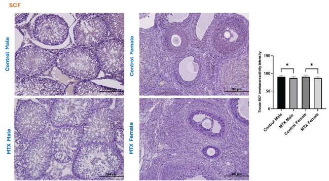
Twenty-four Wistar albino rats, both male and female, were used in our study. Four sets of rats; control male, MTX male, control female, and MTX female were created. The male and female MTX-treated groups received a single intraperitoneal dose of 20 mg/kg MTX. The testes and ovaries of rats sacrificed under general anesthesia were extracted and histopathologically analyzed. In addition, the immunoreactivity intensities of stem cell factor (SCF), mechanistic target of rapamycin (mTOR), and SIRT-1 in both tissues were measured by immunohistochemistry.
Johnsen's testicular biopsy score in the testicular seminiferous tubules was significantly lower in the MTX group than in the control group (p<0.001). The ovary showed substantial follicular degeneration (p<0.05), vascular congestion (p<0.01), and fibrosis (p<0.001). MTX reduced SCF immunoreactivity density in the testis and ovary (p<0.05). Furthermore, MTX reduced mTOR, a marker of autophagy, in the testis (p<0.05) and ovary (p<0.001) compared with the control. SIRT-1 intensity increased dramatically in the testis (p<0.001) and ovary (p<0.01) in the injured group, unlike the mTOR marker.
Our investigation revealed that the gonads incurred significant damage as a result of MTX. One vital option for reducing or eliminating this damage to the ovaries and testicles is the use of anti-oxidant-rich substances.
甲氨蝶呤(MTX)是一种高效的癌症化疗药物。该药物具有性腺毒性作用,主要影响睾丸和卵巢。我们的研究采用组织病理学和免疫组织化学方法,评估使用MTX对睾丸和卵巢组织造成的潜在损害。
我们的研究使用了24只Wistar白化大鼠,雌雄各半。将大鼠分为四组:雄性对照组、雄性MTX组、雌性对照组和雌性MTX组。雄性和雌性MTX处理组均接受单次腹腔注射20mg/kg MTX。在全身麻醉下处死大鼠,取出睾丸和卵巢进行组织病理学分析。此外,通过免疫组织化学测量两种组织中干细胞因子(SCF)、雷帕霉素靶蛋白(mTOR)和SIRT-1的免疫反应强度。
MTX组睾丸生精小管的约翰森睾丸活检评分显著低于对照组(p<0.001)。卵巢出现大量卵泡变性(p<0.05)、血管充血(p<0.01)和纤维化(p<0.001)。MTX降低了睾丸和卵巢中SCF的免疫反应密度(p<0.05)。此外,与对照组相比,MTX降低了睾丸(p<0.05)和卵巢(p<0.001)中自噬标志物mTOR的水平。与mTOR标志物不同,损伤组睾丸(p<0.001)和卵巢(p<0.01)中SIRT-1强度显著增加。
我们的研究表明,MTX对性腺造成了显著损害。减少或消除对卵巢和睾丸这种损害的一个重要选择是使用富含抗氧化剂的物质。