• 文献检索
  • 文档翻译
  • 深度研究
  • 学术资讯
  • Suppr Zotero 插件Zotero 插件
  • 邀请有礼
  • 套餐&价格
  • 历史记录
应用&插件
Suppr Zotero 插件Zotero 插件浏览器插件Mac 客户端Windows 客户端微信小程序
定价
高级版会员购买积分包购买API积分包
服务
文献检索文档翻译深度研究API 文档MCP 服务
关于我们
关于 Suppr公司介绍联系我们用户协议隐私条款
关注我们

Suppr 超能文献

核心技术专利:CN118964589B侵权必究
粤ICP备2023148730 号-1Suppr @ 2026

文献检索

告别复杂PubMed语法,用中文像聊天一样搜索,搜遍4000万医学文献。AI智能推荐,让科研检索更轻松。

立即免费搜索

文件翻译

保留排版,准确专业,支持PDF/Word/PPT等文件格式,支持 12+语言互译。

免费翻译文档

深度研究

AI帮你快速写综述,25分钟生成高质量综述,智能提取关键信息,辅助科研写作。

立即免费体验

130例支气管扩张症患者的胸部X线特征

Chest X-Ray Features in 130 Patients with Bronchiectasis.

作者信息

Sawada Hikaru, Kudoh Ryohei, Yokoyama Atsushi, Hagiwara Akihiko, Hiramatsu Kazufumi, Kadota Jun-Ichi, Komiya Kosaku

机构信息

Respiratory Medicine and Infectious Diseases, Oita University Faculty of Medicine, 1-1 Idaigaoka, Hasama-machi, Yufu, Oita 879-5593, Japan.

Department of Respiratory Medicine, Oita Prefectural Hospital, 2-8-1 Bunyo, Oita 870-8511, Japan.

出版信息

Diseases. 2024 Dec 10;12(12):323. doi: 10.3390/diseases12120323.

DOI:10.3390/diseases12120323
PMID:39727653
原文链接:https://pmc.ncbi.nlm.nih.gov/articles/PMC11726920/
Abstract

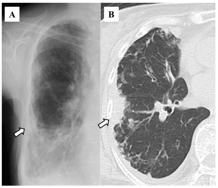
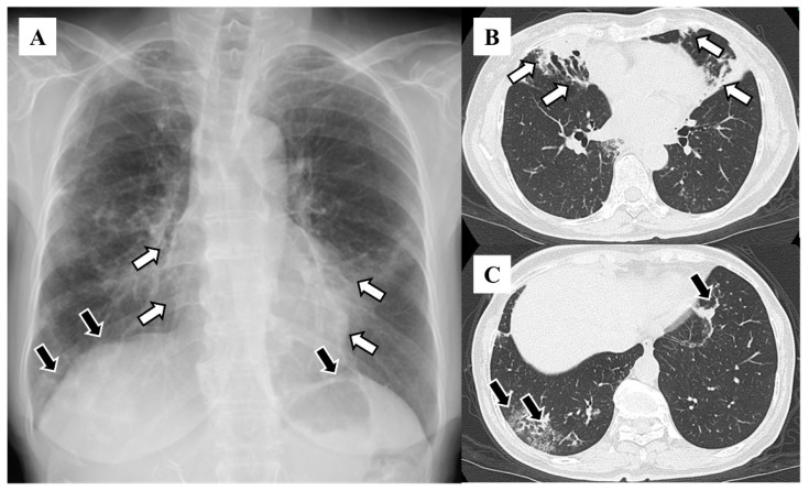
The prevalence of bronchiectasis is increasing globally, and early detection using chest imaging has been encouraged to improve its prognosis. However, the sensitivity of a chest X-ray as a screening tool remains unclear. This study examined the chest X-ray features predictive of bronchiectasis. We retrospectively reviewed the chest X-rays of patients with bronchiectasis diagnosed using high-resolution computed tomography who visited our institute from January 2013 to March 2020. Patients with cardiac pacemakers, lung cancer, and interstitial pneumonia, which might bias the detection of bronchiectasis, were excluded. Two respiratory physicians independently determined the presence or absence of potential features reflecting bronchiectasis, including a vague cardiac silhouette on chest X-rays. The study enrolled 130 patients, including 72 women (55.4%), with a mean age of 72 years. The features observed on chest X-rays included granular shadows (88.5%, n = 115), vague cardiac silhouettes (48.5%, n = 64), nodular shadows (45.4%, n = 59), a tram-track appearance (35.4%, n = 46), pleural thickening (26.9%, n = 35), vague diaphragm silhouettes (25.4%, n = 33), and a ring sign (24.6%, n = 32). The kappa values for these features were 0.271, 0.344, 0.646, 0.256, 0.312, 0.514, and 0.376, respectively. Although traditional chest X-ray features believed to reflect bronchiectasis, such as the tram-track appearance or ring sign, were not frequently seen, vague cardiac silhouettes and granular shadows had high positivity rates, indicating their potential utility for bronchiectasis screening. However, the interobserver concordance rates were unsatisfactory.

摘要

支气管扩张的全球患病率正在上升,因此鼓励采用胸部影像学进行早期检测以改善其预后。然而,胸部X光作为筛查工具的敏感性仍不明确。本研究调查了可预测支气管扩张的胸部X光特征。我们回顾性分析了2013年1月至2020年3月期间到我院就诊、经高分辨率计算机断层扫描诊断为支气管扩张患者的胸部X光片。排除了可能影响支气管扩张检测的心脏起搏器植入患者、肺癌患者和间质性肺炎患者。两名呼吸内科医生独立判断胸部X光片上是否存在反映支气管扩张的潜在特征,包括心脏轮廓模糊。该研究共纳入130例患者,其中女性72例(55.4%),平均年龄72岁。胸部X光片上观察到的特征包括颗粒状阴影(88.5%,n = 115)、心脏轮廓模糊(48.5%,n = 64)、结节状阴影(45.4%,n = 59)、轨道征(35.4%,n = 46)、胸膜增厚(26.9%,n = 35)、膈轮廓模糊(25.4%,n = 33)和环形征(24.6%,n = 32)。这些特征的kappa值分别为0.271、0.344、0.646、0.256、0.312、0.514和0.376。尽管传统上认为反映支气管扩张的胸部X光特征,如轨道征或环形征并不常见,但心脏轮廓模糊和颗粒状阴影的阳性率较高,表明它们在支气管扩张筛查中具有潜在用途。然而观察者间的一致性率并不理想。

https://cdn.ncbi.nlm.nih.gov/pmc/blobs/a35d/11726920/238cdaff442e/diseases-12-00323-g004.jpg
https://cdn.ncbi.nlm.nih.gov/pmc/blobs/a35d/11726920/73326cd0c077/diseases-12-00323-g001.jpg
https://cdn.ncbi.nlm.nih.gov/pmc/blobs/a35d/11726920/1a0f58bd457c/diseases-12-00323-g002.jpg
https://cdn.ncbi.nlm.nih.gov/pmc/blobs/a35d/11726920/a3ab5d734966/diseases-12-00323-g003.jpg
https://cdn.ncbi.nlm.nih.gov/pmc/blobs/a35d/11726920/238cdaff442e/diseases-12-00323-g004.jpg

相似文献

1
Chest X-Ray Features in 130 Patients with Bronchiectasis.130例支气管扩张症患者的胸部X线特征
Diseases. 2024 Dec 10;12(12):323. doi: 10.3390/diseases12120323.
2
Could automated analysis of chest X-rays detect early bronchiectasis in children?胸部 X 光片的自动化分析能否检测出儿童早期的支气管扩张症?
Eur J Pediatr. 2021 Oct;180(10):3171-3179. doi: 10.1007/s00431-021-04061-8. Epub 2021 Apr 28.
3
Clinical value and radiographic features of low dose CT scans compared to X rays in diagnosing mycoplasma pneumonia in children.低剂量CT扫描与X线相比在诊断儿童支原体肺炎中的临床价值及影像学特征
Sci Rep. 2025 Mar 17;15(1):9162. doi: 10.1038/s41598-025-94006-0.
4
Comparison of the chest computed tomography findings between patients with pulmonary tuberculosis and those with Mycobacterium avium complex lung disease.肺结核患者与鸟分枝杆菌复合群肺病患者胸部计算机断层扫描结果的比较。
Respir Investig. 2020 May;58(3):137-143. doi: 10.1016/j.resinv.2019.12.006. Epub 2020 Feb 23.
5
Chest CT-based Assessment of 1-year Outcomes after Moderate COVID-19 Pneumonia.基于胸部 CT 的中度 COVID-19 肺炎 1 年后结局评估。
Radiology. 2022 Nov;305(2):479-485. doi: 10.1148/radiol.220019. Epub 2022 May 10.
6
CT scans of the chest in carriers of human T-cell lymphotropic virus type 1: presence of interstitial pneumonia.胸部 CT 扫描在人 T 细胞嗜淋巴细胞病毒 1 携带者中的表现:间质性肺炎。
Acad Radiol. 2012 Aug;19(8):952-7. doi: 10.1016/j.acra.2012.03.020. Epub 2012 May 10.
7
Radiologist agreement on the quantification of bronchiectasis by high-resolution computed tomography.放射科医生对高分辨率计算机断层扫描定量支气管扩张的一致性。
Radiol Bras. 2017 Jan-Feb;50(1):26-31. doi: 10.1590/0100-3984.2015.0146.
8
Radiologic types of Mycobacterium xenopi pulmonary disease: different patients with similar short-term outcomes.分枝杆菌肺病的放射学类型:不同患者具有相似的短期结局。
Eur J Clin Microbiol Infect Dis. 2019 Feb;38(2):373-381. doi: 10.1007/s10096-018-3437-x. Epub 2018 Dec 14.
9
Thoracic imaging tests for the diagnosis of COVID-19.用于 COVID-19 诊断的胸部影像学检查。
Cochrane Database Syst Rev. 2022 May 16;5(5):CD013639. doi: 10.1002/14651858.CD013639.pub5.
10
Chest Computed Tomography Features of Nontuberculous Mycobacterial Pulmonary Disease Versus Asymptomatic Colonization: A Cross-sectional Cohort Study.非结核分枝杆菌肺病与无症状定植的胸部计算机断层扫描特征:一项横断面队列研究
J Thorac Imaging. 2022 May 1;37(3):140-145. doi: 10.1097/RTI.0000000000000610. Epub 2021 Jul 21.

本文引用的文献

1
CT Imaging Assessment of Response to Treatment in Allergic Bronchopulmonary Aspergillosis in Adults With Bronchial Asthma.CT 影像学在哮喘成人变应性支气管肺曲霉病治疗反应评估中的作用。
Chest. 2024 Jun;165(6):1307-1318. doi: 10.1016/j.chest.2024.02.026. Epub 2024 Feb 20.
2
Bronchiectasis in Europe: data on disease characteristics from the European Bronchiectasis registry (EMBARC).欧洲支气管扩张症:欧洲支气管扩张症注册研究(EMBARC)中有关疾病特征的数据。
Lancet Respir Med. 2023 Jul;11(7):637-649. doi: 10.1016/S2213-2600(23)00093-0. Epub 2023 Apr 24.
3
Bronchiectasis - A Clinical Review.
支气管扩张症——临床综述
N Engl J Med. 2022 Aug 11;387(6):533-545. doi: 10.1056/NEJMra2202819.
4
Diagnosis and Management of Idiopathic Pulmonary Fibrosis.特发性肺纤维化的诊断与管理。
R I Med J (2013). 2021 Sep 1;104(7):26-29.
5
British Thoracic Society guideline for bronchiectasis in adults.英国胸科学会成人支气管扩张指南。
BMJ Open Respir Res. 2018 Dec 28;5(1):e000348. doi: 10.1136/bmjresp-2018-000348. eCollection 2018.
6
Adult Patients With Bronchiectasis: A First Look at the US Bronchiectasis Research Registry.成年支气管扩张症患者:美国支气管扩张症研究注册库的初步观察
Chest. 2017 May;151(5):982-992. doi: 10.1016/j.chest.2016.10.055. Epub 2016 Nov 23.
7
Changes in the incidence, prevalence and mortality of bronchiectasis in the UK from 2004 to 2013: a population-based cohort study.2004年至2013年英国支气管扩张症的发病率、患病率及死亡率变化:一项基于人群的队列研究。
Eur Respir J. 2016 Jan;47(1):186-93. doi: 10.1183/13993003.01033-2015. Epub 2015 Nov 5.
8
Lung abscess-etiology, diagnostic and treatment options.肺脓肿的病因、诊断和治疗选择。
Ann Transl Med. 2015 Aug;3(13):183. doi: 10.3978/j.issn.2305-5839.2015.07.08.
9
An analysis of etiology, causal pathogens, imaging patterns, and treatment of Japanese patients with bronchiectasis.日本支气管扩张症患者的病因、致病病原体、影像学表现及治疗分析
Respir Investig. 2015 Jan;53(1):37-44. doi: 10.1016/j.resinv.2014.09.004. Epub 2014 Nov 7.
10
Multidimensional approach to non-cystic fibrosis bronchiectasis: the FACED score.多维方法治疗非囊性纤维化性支气管扩张症:FACED 评分。
Eur Respir J. 2014 May;43(5):1357-67. doi: 10.1183/09031936.00026313. Epub 2013 Nov 14.