Cao Wenli, Pan Xiaofeng, Jin Liming, Liu Jie, Cao Jie, Jin Lei, Wei Fangqiang
Department of General Surgery, Cancer center, Division of Hepatobiliary and Pancreatic Surgery, Zhejiang Provincial People's Hospital, Affiliated People's Hospital, Hangzhou Medical College, Hangzhou, Zhejiang Province, China.
Department of Public Health, Hangzhou Medical College, Hangzhou, Zhejiang Province, China.
PLoS One. 2024 Dec 27;19(12):e0316199. doi: 10.1371/journal.pone.0316199. eCollection 2024.
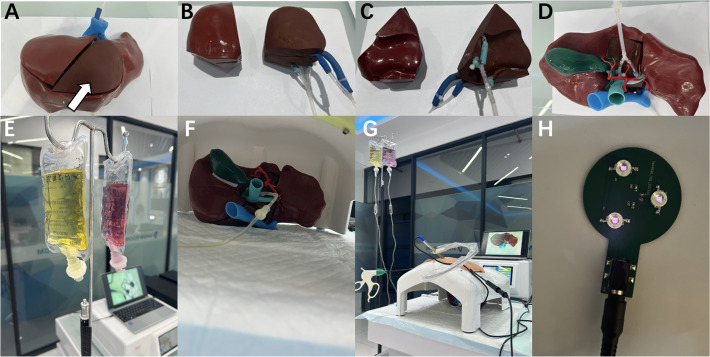
Complex liver cancer is often difficult to expose or dissect, and the surgery is often challenging. 3D-printed models may realistically present 3D anatomical structure, which has certain value in planning and training of liver surgery. However, the existing 3D-printed models are all monolithic models, which are difficult to reuse and limited in clinical application. It is also rare to carry fluorescence to accurately present tumor lesions. Here we report reusable fluorescent assembled 3D-printed models to mimic minimally invasive resection of complex liver cancer. Based on the models, multiple copies of liver lesion structure assembled accessories can be printed for the same patient or different patients, ensuring the quantity and quality of simulated surgical training, and greatly reducing the cost of simulated surgical training. The addition of fluorescence is helpful in accurately presenting tumor lesions. The reusable fluorescent assembled 3D-printed models may mimic minimally invasive resection of complex liver cancer, demonstrating potential value in simulated surgery.
复杂肝癌往往难以暴露或解剖,手术通常具有挑战性。3D打印模型可以逼真地呈现三维解剖结构,这在肝脏手术的规划和训练中具有一定价值。然而,现有的3D打印模型都是整体模型,难以重复使用且临床应用受限。携带荧光以准确呈现肿瘤病变的情况也很少见。在此,我们报告可重复使用的荧光组装3D打印模型,以模拟复杂肝癌的微创切除。基于这些模型,可以为同一患者或不同患者打印多个肝脏病变结构组装配件副本,确保模拟手术训练的数量和质量,并大大降低模拟手术训练的成本。添加荧光有助于准确呈现肿瘤病变。可重复使用的荧光组装3D打印模型可以模拟复杂肝癌的微创切除,在模拟手术中显示出潜在价值。