Kros Johan M, Zeneyedpour Lona, Pedrosa Rute M S M, Belcaid Zineb, Dik Willem A, Luider Theo M, Mustafa Dana A M
Department of Pathology, The Tumor Immuno-Pathology Laboratory, Erasmus University Medical Center, Wytemaweg 80, 3000 DR, Rotterdam, The Netherlands.
Department of Neurology, Laboratory of Neuro‑Oncology, Clinical and Cancer Proteomics, Erasmus University Medical Center, Wytemaweg 80, 3000 DR, Rotterdam, The Netherlands.
Sci Rep. 2024 Dec 28;14(1):31516. doi: 10.1038/s41598-024-83301-x.
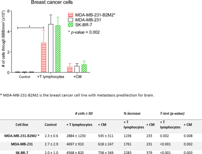
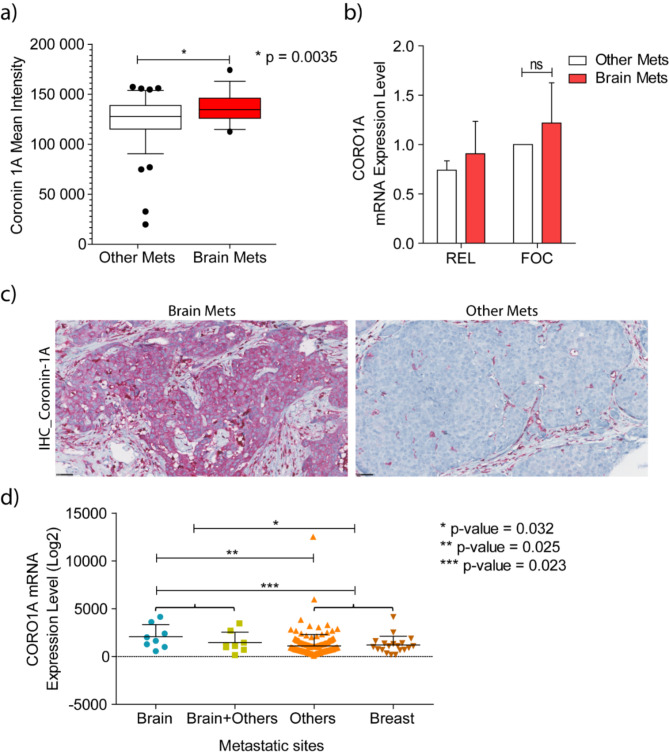
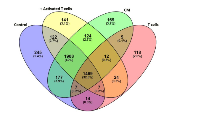
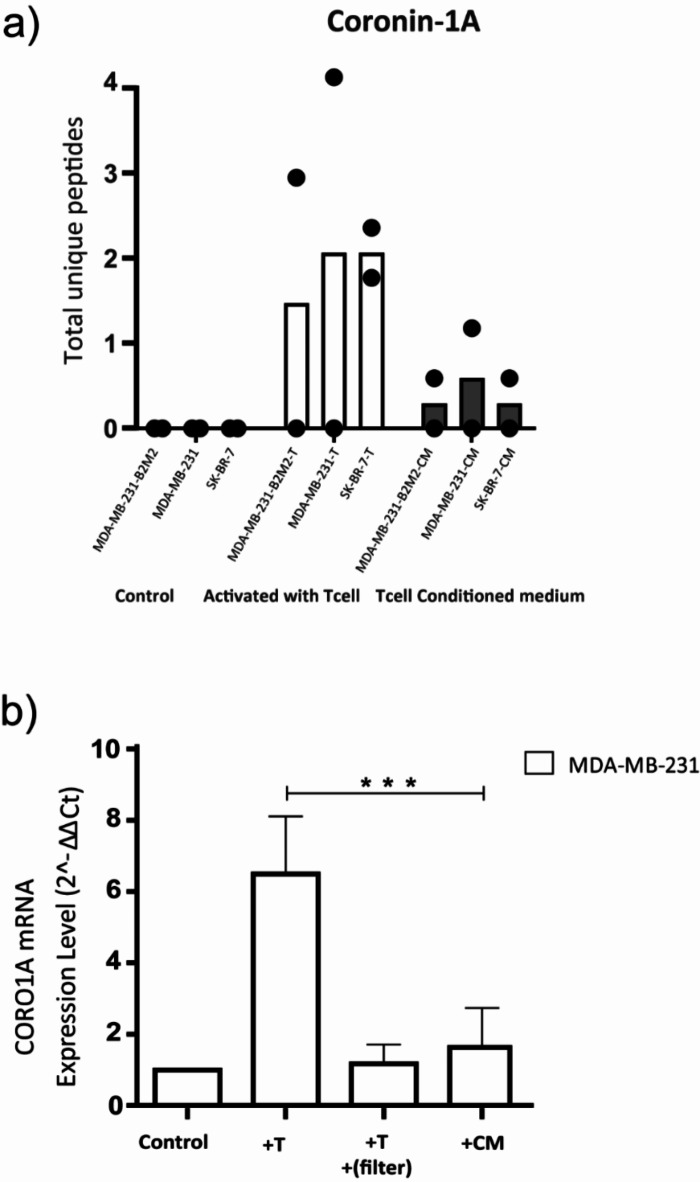
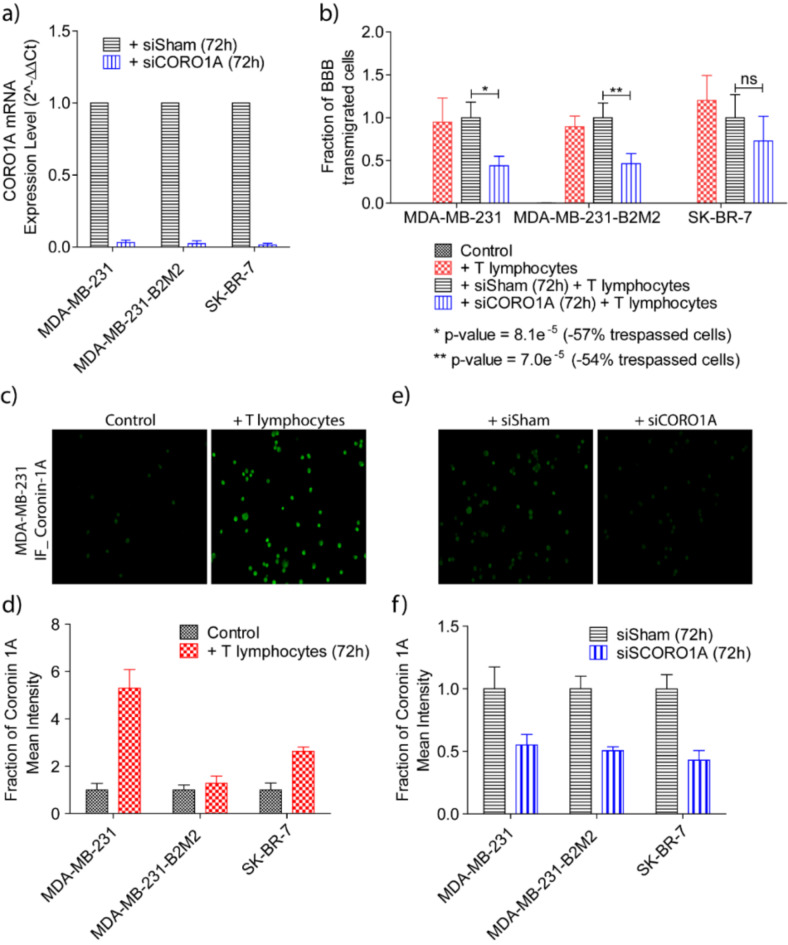
In previous work we discovered that T lymphocytes play a prominent role in the rise of brain metastases of ER-negative breast cancers. In the present study we explored how T lymphocytes promote breast cancer cell penetration through the blood brain barrier (BBB). An in vitro BBB model was employed to study the effects of T lymphocytes on BBB trespassing capacity of three different breast carcinoma cell lines. Differential protein expression was explored by comparing the proteomes of the breast cancer cells before and after co-culture with activated T lymphocytes using liquid chromatography-mass spectrometry (LC-MS). siRNA was used to silence protein expression in the breast cancer cells to study contribution to in vitro BBB passage. Furthermore, protein expression in primary breast cancer tissues was explored and related to brain-metastatic potential. Co-culturing with activated T lymphocytes or their conditioned medium (CM) resulted in increased passage through the in vitro BBB. The effects were less for cell line MDA-MB-231-B2M2 (brain affinity) as compared to MDA-MB-231 and SK-BR-7. Mass spectrometry-based proteomics revealed significant alterations in the expression of 35 proteins by the breast cancer cell lines upon T cell contact. Among the proteins is coronin-1 A, a protein related to cell motility. Knockdown of CORO1A in the breast cancer cells reduced their ability to cross the artificial BBB to 60%. The effects were significantly less for the cell line derived from breast cancer with affinity for brain. The expression of coronin-1A was confirmed by immunohistochemistry and RT-PCR of 52 breast cancer samples of patients with metastasized breast cancers, with and without brain locations. Lastly, CORO1A upregulation was validated in a publicly available mRNA expression database from 204 primary breast cancers with known metastatic sites. We conclude that T lymphocytes trigger cancer cells to express proteins including coronin-1A that enable the cancer cells to cross an in vitro BBB. In addition, a prominent role of coronin-1A in the formation of cerebral metastases in breast cancer patients is strongly suggestive by its upregulation in tissue samples of breast cancer patients with brain metastases.
在之前的研究中,我们发现T淋巴细胞在雌激素受体阴性乳腺癌脑转移的发生过程中发挥着重要作用。在本研究中,我们探讨了T淋巴细胞如何促进乳腺癌细胞穿透血脑屏障(BBB)。采用体外血脑屏障模型研究T淋巴细胞对三种不同乳腺癌细胞系穿越血脑屏障能力的影响。通过液相色谱-质谱联用(LC-MS)比较乳腺癌细胞与活化T淋巴细胞共培养前后的蛋白质组,探索差异蛋白表达。使用小干扰RNA(siRNA)沉默乳腺癌细胞中的蛋白表达,以研究其对体外血脑屏障穿越的作用。此外,还探索了原发性乳腺癌组织中的蛋白表达,并将其与脑转移潜能相关联。与活化T淋巴细胞或其条件培养基(CM)共培养导致体外血脑屏障的穿越增加。与MDA-MB-231和SK-BR-7相比,细胞系MDA-MB-231-B2M2(脑亲和性)的这种作用较小。基于质谱的蛋白质组学显示,T细胞接触后,乳腺癌细胞系中35种蛋白质的表达发生了显著变化。其中包括冠蛋白-1A,一种与细胞运动相关的蛋白质。敲低乳腺癌细胞中的CORO1A可将其穿越人工血脑屏障的能力降低至60%。对于源自具有脑亲和性的乳腺癌的细胞系,这种作用明显较小。通过对52例有或无脑转移的转移性乳腺癌患者的乳腺癌样本进行免疫组织化学和逆转录-聚合酶链反应(RT-PCR),证实了冠蛋白-1A的表达。最后,在一个来自204例已知转移部位的原发性乳腺癌的公开可用mRNA表达数据库中验证了CORO1A的上调。我们得出结论,T淋巴细胞触发癌细胞表达包括冠蛋白-1A在内的蛋白质,使癌细胞能够穿越体外血脑屏障。此外,冠蛋白-1A在乳腺癌患者脑转移形成中的重要作用在有脑转移的乳腺癌患者组织样本中的上调中得到了有力提示。