Wang Haohao, Jiang Rendong, Dong Zhishang, Zhao Dongyue, Zhao Jianli, Shi Chao, Yuan Zhen
Department of Orthopedics, The First Affiliated Hospital of Shandong First Medical University & Shandong Provincial Qianfoshan Hospital, Jinan, Shandong, China.
Front Surg. 2024 Dec 13;11:1487156. doi: 10.3389/fsurg.2024.1487156. eCollection 2024.
Hemophilic arthritis (HA) is associated with significant changes in the morphology of mature knee joints due to abnormal growth plate development. Previous studies have established marked distinctions between the femur and tibia of subjects with Haemophilia and those with osteoarthritis (OA). This study explored the morphological characteristics of the patella and patellofemoral joint in subjects with Haemophilia. These findings can inform the design of knee joint prostheses tailored to this condition, improve the precision of total knee replacement surgery, and reduce postoperative knee pain and patellar dislocation.
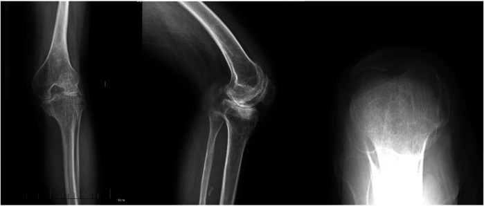
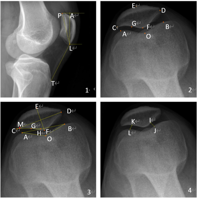
Before surgery, we conducted preoperative measurements of patellar length, patellar diagonal length, patellar ligament length, patellar width, patellar thickness, the INSALL index, the lateral patellofemoral angle, the trochlear groove angle,the patellar lateral displacement rate, and the patellofemoral index using lateral and axial x-ray images in 40 subjects with Haemophilia, 40 OA patients, and 40 normal individuals.
Significant statistical differences in certain morphological parameters were observed among the three groups of patients ( < 0.05). Compared with the OA and normal control groups, the HA group presented significant disparities in patellar thickness, patellar ligament length, the Insall ratio, the patellar lateral shift rate, the lateral patellar angle, and the patellofemoral index.
Compared with OA and normal individuals, Subjects with Haemophilia presented with smaller and thinner patellae, more significant patellar ligament contracture, reduced patellar height, and more pronounced patellar dislocation. Consequently, during total knee arthroplasty, we lean toward patellar reshaping in subjects with Haemophilia, exercise caution when considering patellar replacement, and, for those with severe preoperative patellar dislocation, perform intraoperative lateral retinacular release.
血友病性关节炎(HA)由于生长板发育异常,与成熟膝关节的形态发生显著变化有关。先前的研究已经明确了血友病患者与骨关节炎(OA)患者在股骨和胫骨方面的明显差异。本研究探讨了血友病患者髌骨和髌股关节的形态特征。这些发现可为针对这种情况的膝关节假体设计提供参考,提高全膝关节置换手术的精度,并减少术后膝关节疼痛和髌骨脱位。
在手术前,我们使用侧位和轴位X线图像,对40例血友病患者、40例OA患者和40例正常个体进行了髌骨长度、髌骨对角线长度、髌韧带长度、髌骨宽度、髌骨厚度、Insall指数、髌股外侧角、滑车沟角、髌骨外侧移位率和髌股指数的术前测量。
三组患者在某些形态学参数上存在显著统计学差异(<0.05)。与OA组和正常对照组相比,HA组在髌骨厚度、髌韧带长度、Insall比率、髌骨外侧移位率、髌骨外侧角和髌股指数方面存在显著差异。
与OA患者和正常个体相比,血友病患者的髌骨更小、更薄,髌韧带挛缩更明显,髌骨高度降低,髌骨脱位更显著。因此,在全膝关节置换术中,对于血友病患者,我们倾向于进行髌骨重塑,在考虑髌骨置换时要谨慎,对于术前髌骨脱位严重的患者,术中进行外侧支持带松解。