Ito Eisaku, Ohki Takao, Toya Naoki, Naganuma Hirokuni, Kawada Noriyasu, Muramatsu Koichi, Fukasawa Nei, Miyake Misayo, Maeda Miku, Shimoda Masayuki
Division of Vascular Surgery, Department of Surgery, The Jikei University Kashiwa Hospital, Chiba, Japan.
Division of Vascular Surgery, Department of Surgery, The Jikei University School of Medicine, Tokyo, Japan.
Aorta (Stamford). 2024 Apr;12(2):25-31. doi: 10.1055/s-0044-1791669. Epub 2024 Dec 31.
Aortic wall enhancement (AWE), evaluated with computed tomography angiography in Type B aortic dissection, is associated with aortic remodeling. This study aimed to evaluate the relationship between AWE and pathological findings of the aortic wall using an aortic wall sample from a Type A aortic dissection (TAD).
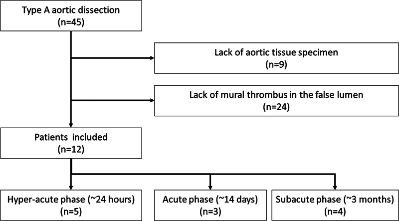
We examined patients with TAD treated between January 2012 and February 2023.
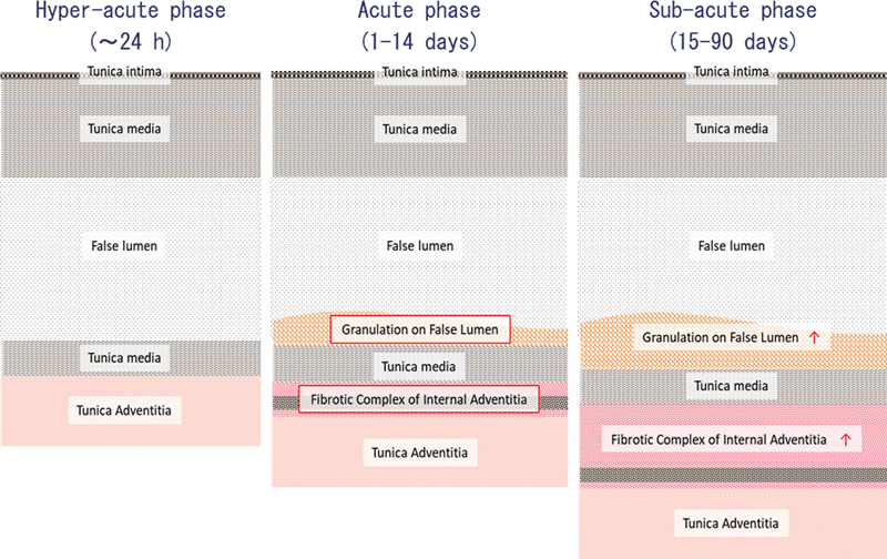
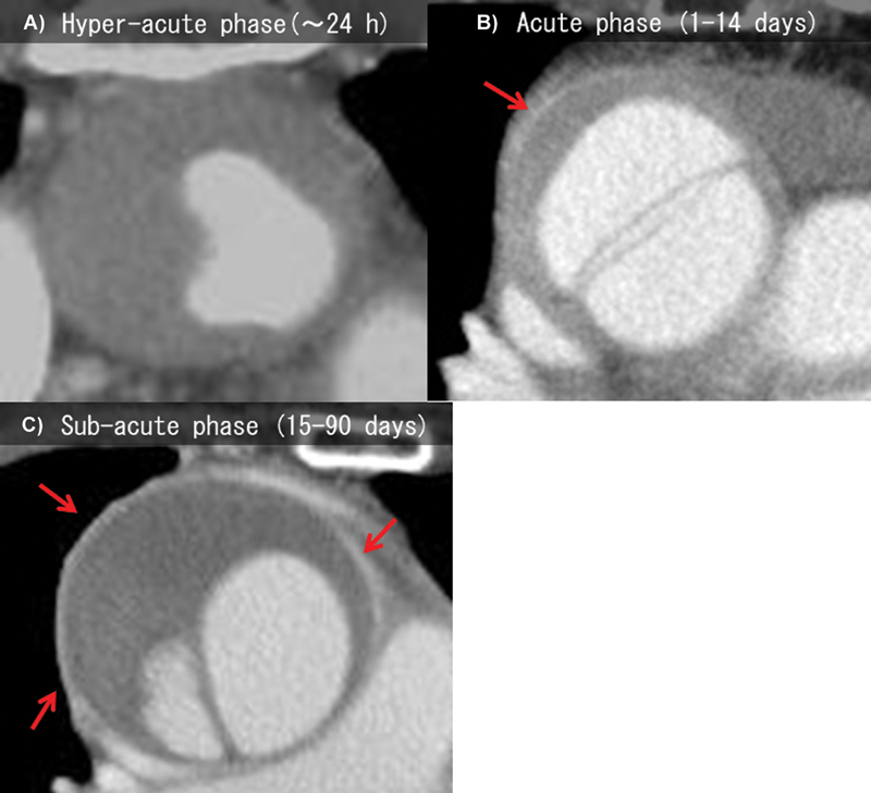
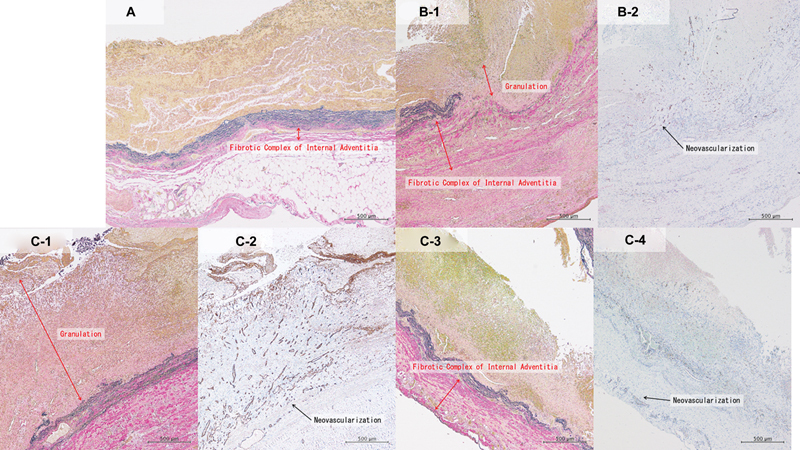
Twelve cases were examined pathologically: five cases in the hyperacute phase, three in the acute phase, and four in the subacute phase. AWE thickness significantly differed as time progressed (0 vs. 1.7 vs. 2.8 mm, < 0.001). A significant increase in granulation was observed in the acute and subacute phases (0 vs. 761 vs. 423 µm, < 0.001). Furthermore, a fibrous complex of internal adventitia (FCIA) developed on the medial side of the adventitia over time since its onset and was found to be thickened (175 vs. 415 vs. 1,078 µm, < 0.001). The thickness of the granulation tissue and FCIA, where there was abundant neovascularization, was consistent with the thickness of the AWE.
AWE was observed in TAD and increased as time progressed. FCIA and granulation tissue developed, and AWE reflected neovascularization at the adventitia.
在B型主动脉夹层中,通过计算机断层血管造影评估的主动脉壁强化(AWE)与主动脉重塑有关。本研究旨在使用A型主动脉夹层(TAD)的主动脉壁样本评估AWE与主动脉壁病理结果之间的关系。
我们检查了2012年1月至2023年2月期间接受治疗的TAD患者。
对12例患者进行了病理检查:超急性期5例,急性期3例,亚急性期4例。随着时间的推移,AWE厚度有显著差异(0 vs. 1.7 vs. 2.8毫米,<0.001)。在急性期和亚急性期观察到肉芽组织显著增加(0 vs. 761 vs. 423微米,<0.001)。此外,自发病以来,外膜内侧逐渐形成内膜外膜纤维复合体(FCIA),且发现其增厚(175 vs. 415 vs. 1078微米,<0.001)。肉芽组织和FCIA的厚度,即新生血管丰富的部位,与AWE的厚度一致。
在TAD中观察到AWE,并随时间推移而增加。FCIA和肉芽组织形成,且AWE反映了外膜的新生血管形成。