Zhong Ruiqi, Zhang Ying, Qiu Wenzhuo, Zhang Kaipeng, Feng Qianqian, Cao Xiuxue, Huang Qixin, Zhang Yijing, Guo Yuanyuan, Guo Jia, Zhao Lingyu, Wang Xiuhong, Wang Shuhao, Cui Lifang, Wang Aimin, Qian Haili, Ma Fei
4+4 Medical Doctor Program, Chinese Academy of Medical Sciences & Peking Union Medical College, Beijing 100730, China.
Department of Medical Oncology, National Cancer Center/National Clinical Research Center for Cancer/Cancer Hospital, Chinese Academy of Medical Sciences and Peking Union Medical College, Beijing 100021, China.
Int J Biol Sci. 2025 Jan 1;21(1):363-381. doi: 10.7150/ijbs.102744. eCollection 2025.
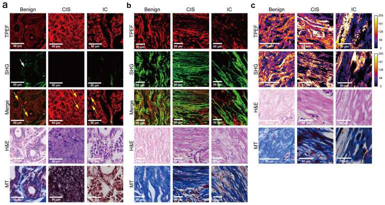
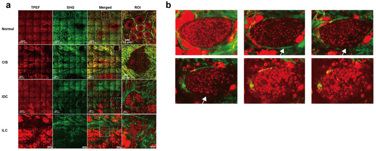
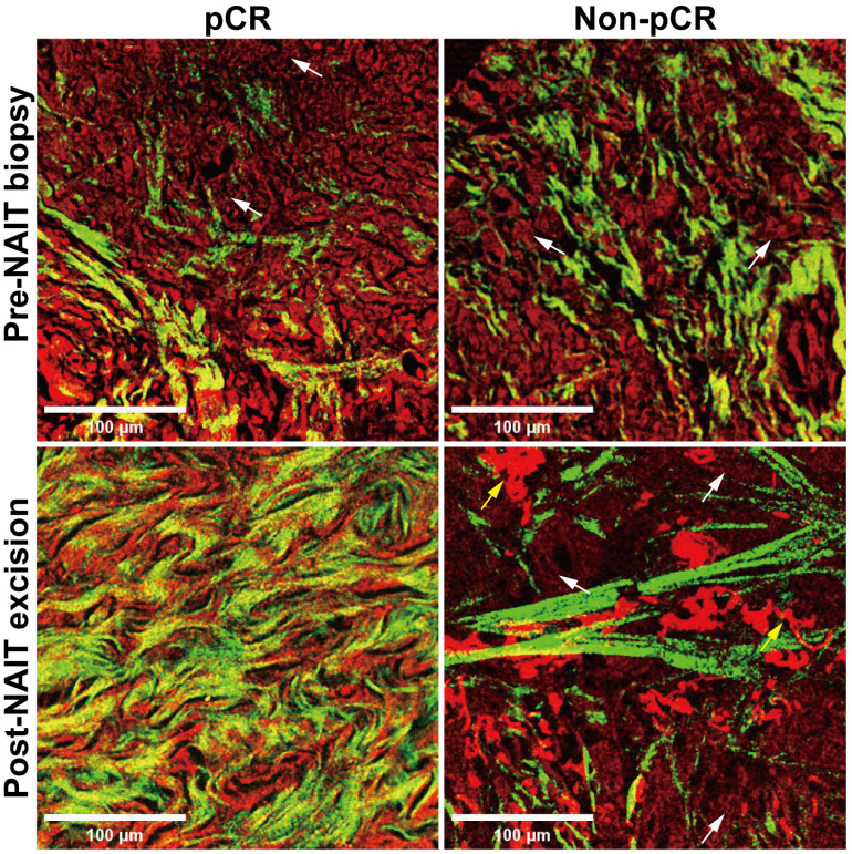
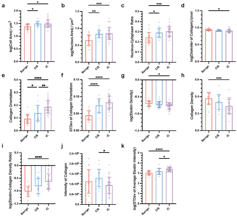
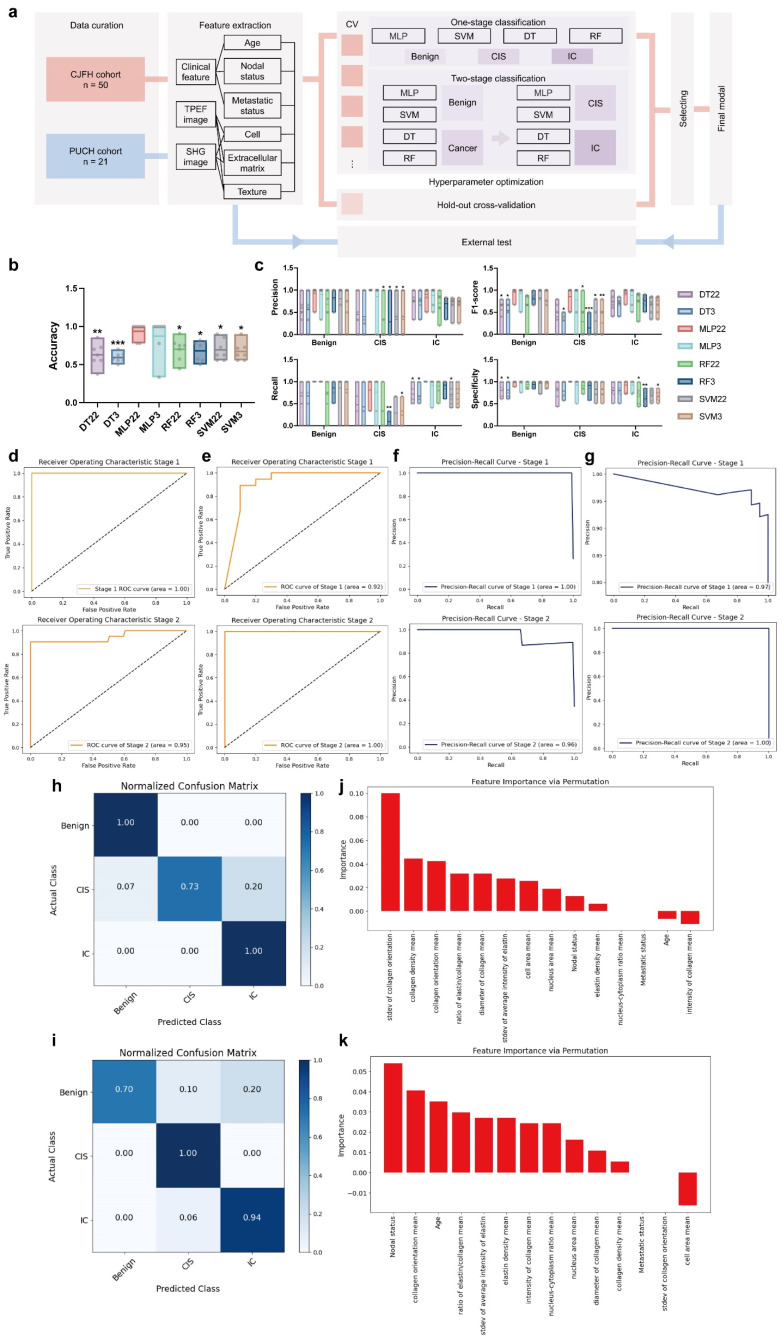
Accurate diagnosis and assessment of breast cancer treatment responses are critical challenges in clinical practice, influencing patient treatment strategies and ultimately long-term prognosis. Currently, diagnosing breast cancer and evaluating the efficacy of neoadjuvant immunotherapy (NAIT) primarily rely on pathological identification of tumor cell morphology, count, and arrangement. However, when tumors are small, the tumors and tumor beds are difficult to detect; relying solely on tumor cell identification may lead to false negatives. In this study, we used the label-free multiphoton microscopy (MPM) method to quantitatively analyze breast tissue at the cellular, extracellular, and textural levels, and identified 11 key factors that can effectively distinguish different types of breast diseases. Key factors and clinical data are used to train a two-stage machine learning automatic diagnosis model, MINT, to accurately diagnose breast cancer. The classification capability of MINT was validated in independent cohorts (stage 1 AUC = 0.92; stage 2 AUC = 1.00). Furthermore, we also found that some factors could predict and assess the efficacy of NAIT, demonstrating the potential of label-free MPM in breast cancer diagnosis and treatment. We envision that in the future, label-free MPM can be used to complement stromal and textural information in pathological tissue, benefiting breast cancer diagnosis and neoadjuvant therapy efficacy prediction, thereby assisting clinicians in formulating personalized treatment plans.
准确诊断和评估乳腺癌治疗反应是临床实践中的关键挑战,影响患者的治疗策略及最终的长期预后。目前,诊断乳腺癌和评估新辅助免疫疗法(NAIT)的疗效主要依赖于肿瘤细胞形态、数量及排列的病理鉴定。然而,当肿瘤较小时,肿瘤及其瘤床难以检测;仅依靠肿瘤细胞鉴定可能导致假阴性。在本研究中,我们使用无标记多光子显微镜(MPM)方法在细胞、细胞外和组织纹理水平对乳腺组织进行定量分析,并确定了11个可有效区分不同类型乳腺疾病的关键因素。利用关键因素和临床数据训练一个两阶段机器学习自动诊断模型MINT,以准确诊断乳腺癌。MINT的分类能力在独立队列中得到验证(第一阶段AUC = 0.92;第二阶段AUC = 1.00)。此外,我们还发现一些因素可预测和评估NAIT的疗效,证明了无标记MPM在乳腺癌诊断和治疗中的潜力。我们设想,未来无标记MPM可用于补充病理组织中的基质和组织纹理信息,有助于乳腺癌诊断和新辅助治疗疗效预测,从而协助临床医生制定个性化治疗方案。