Wang Luwei, Chen Yue, Guo Jiaqing, Weng Xiaoyu, Yan Wei, Song Jun, Ye Tong, Qu Junle
Center for Biomedical Optics and Photonics & College of Physics and Optoelectronic Engineering, Key Laboratory of Optoelectronic Devices and Systems of Ministry of Education and Guangdong Province, Shenzhen University, Shenzhen, 518060, China.
The Photonics Center of Shenzhen University, Shenzhen University, Shenzhen, 518060, China.
Light Sci Appl. 2025 Jan 3;14(1):32. doi: 10.1038/s41377-024-01711-y.
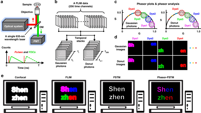
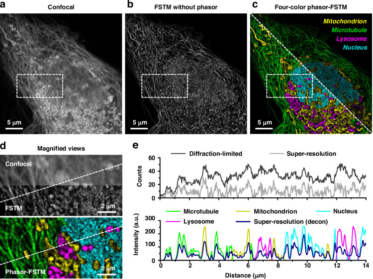
Multicolor microscopy and super-resolution optical microscopy are two widely used techniques that greatly enhance the ability to distinguish and resolve structures in cellular imaging. These methods have individually transformed cellular imaging by allowing detailed visualization of cellular and subcellular structures, as well as organelle interactions. However, integrating multicolor and super-resolution microscopy into a single method remains challenging due to issues like spectral overlap, crosstalk, photobleaching, phototoxicity, and technical complexity. These challenges arise from the conflicting requirements of using different fluorophores for multicolor labeling and fluorophores with specific properties for super-resolution imaging. We propose a novel multicolor super-resolution imaging method called phasor-based fluorescence spatiotemporal modulation (Phasor-FSTM). This method uses time-resolved detection to acquire spatiotemporal data from encoded photons, employs phasor analysis to simultaneously separate multiple components, and applies fluorescence modulation to create super-resolution images. Phasor-FSTM enables the identification of multiple structural components with greater spatial accuracy on an enhanced laser scanning confocal microscope using a single-wavelength laser. To demonstrate the capabilities of Phasor-FSTM, we performed two-color to four-color super-resolution imaging at a resolution of ~λ/5 and observed the interactions of organelles in live cells during continuous imaging for a duration of over 20 min. Our method stands out for its simplicity and adaptability, seamlessly fitting into existing laser scanning microscopes without requiring multiple laser lines for excitation, which also provides a new avenue for other super-resolution imaging technologies based on different principles to build multi-color imaging systems with the requirement of a lower budget.
多色显微镜和超分辨率光学显微镜是两种广泛使用的技术,它们极大地增强了在细胞成像中区分和分辨结构的能力。这些方法通过允许对细胞和亚细胞结构以及细胞器相互作用进行详细可视化,各自改变了细胞成像。然而,由于光谱重叠、串扰、光漂白、光毒性和技术复杂性等问题,将多色和超分辨率显微镜集成到单一方法中仍然具有挑战性。这些挑战源于多色标记使用不同荧光团以及超分辨率成像使用具有特定特性的荧光团的相互冲突的要求。我们提出了一种名为基于相量的荧光时空调制(Phasor-FSTM)的新型多色超分辨率成像方法。该方法使用时间分辨检测从编码光子获取时空数据,采用相量分析同时分离多个成分,并应用荧光调制来创建超分辨率图像。Phasor-FSTM能够在增强型激光扫描共聚焦显微镜上使用单波长激光以更高的空间精度识别多个结构成分。为了证明Phasor-FSTM的能力,我们以~λ/5的分辨率进行了双色到四色超分辨率成像,并在连续成像超过20分钟的过程中观察了活细胞中细胞器的相互作用。我们的方法因其简单性和适应性而脱颖而出,无需多条激光线进行激发即可无缝融入现有的激光扫描显微镜,这也为基于不同原理的其他超分辨率成像技术构建低成本的多色成像系统提供了一条新途径。