Espinosa Gabriel, Salinas-Varas Constanza, Rojas-Barón Lisbeth, Preußer Christian, Pogge von Strandmann Elke, Gärtner Ulrich, Conejeros Iván, Hermosilla Carlos, Taubert Anja
Institute of Parasitology, Justus Liebig University Giessen, Giessen, Germany.
Core Facility Extracellular Vesicles, Center for Tumor Biology and Immunology, Philipps University of Marburg, Marburg, Germany.
Front Immunol. 2024 Dec 19;15:1509355. doi: 10.3389/fimmu.2024.1509355. eCollection 2024.
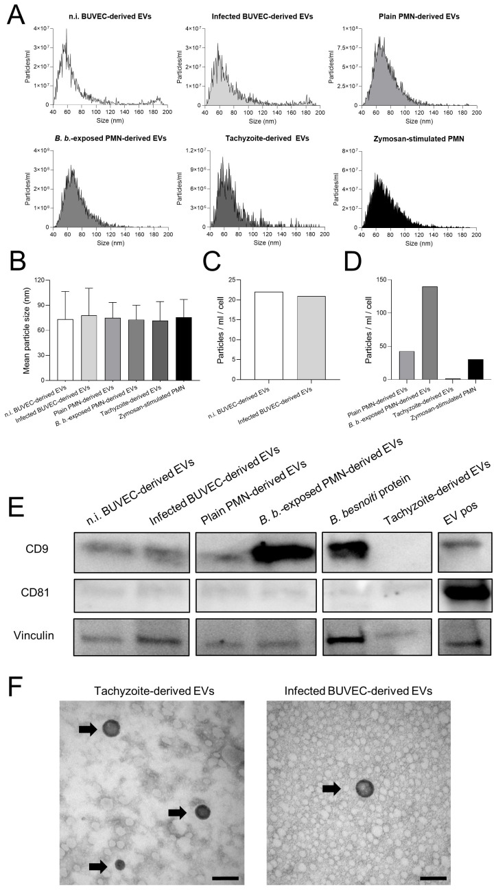
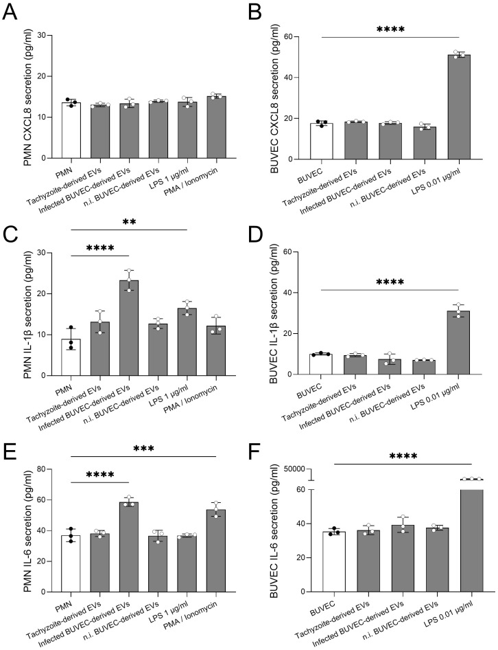
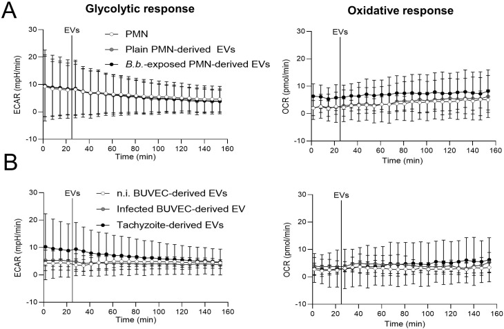
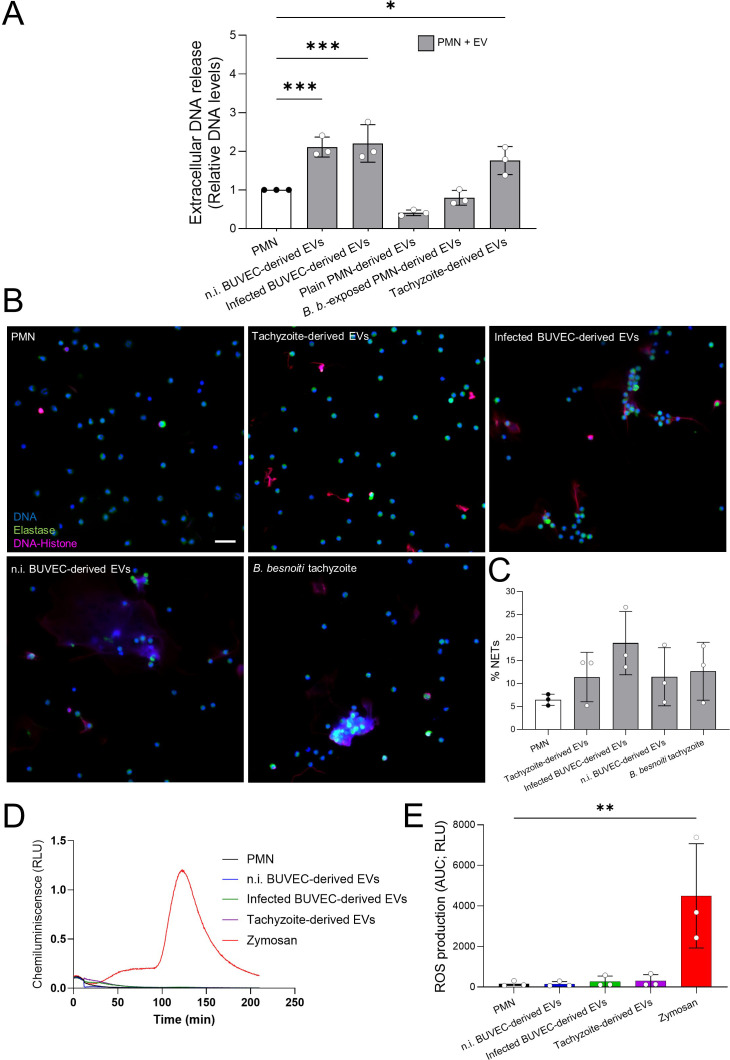
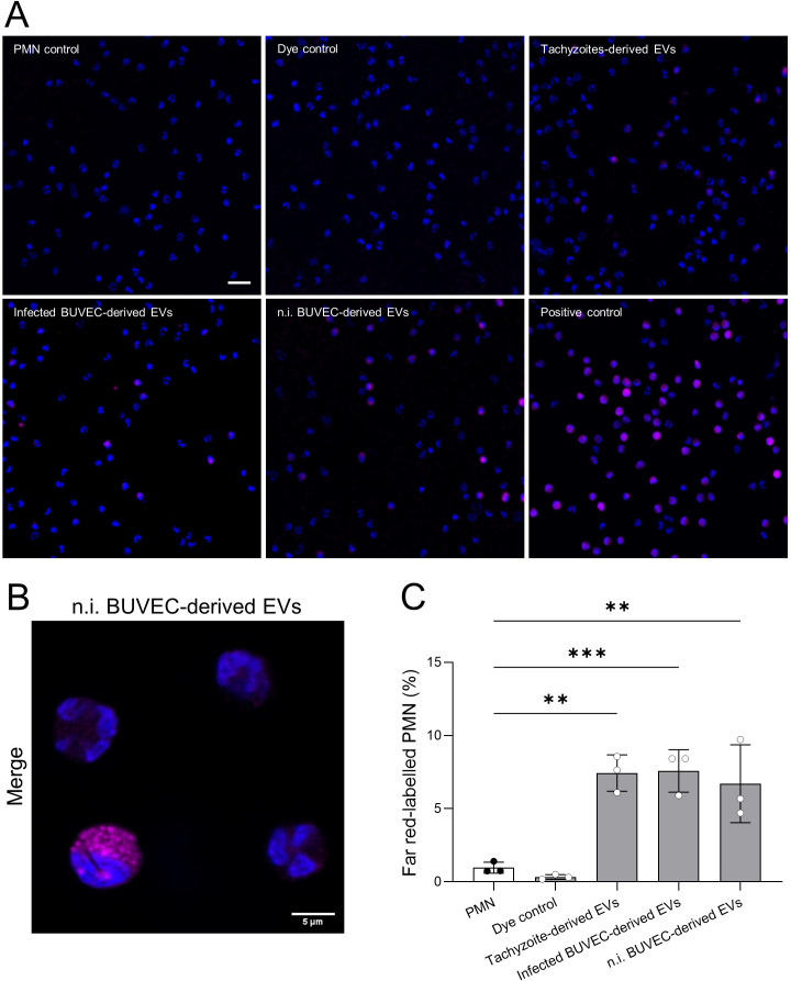
Bovine besnoitiosis is a re-emerging cattle disease caused by the apicomplexan parasite , which severely affects individual animal welfare and profitability in cattle industry. We recently showed that tachyzoite exposure to bovine polymorphonuclear neutrophils (PMN) effectively triggers neutrophil extracellular trap (NET) formation, leading to parasite immobilization hampering host cell infection. So far, the triggers of this defense mechanism remain unclear. Emerging evidence indicates that extracellular vesicles (EVs) modulate PMN effector functions, such as ROS production or NET formation. Therefore, we tested whether exposure of bovine PMN to EVs from different cellular sources affects classical PMN effector functions and cytokine/chemokine secretion. EVs were isolated from -infected and non-infected host cells (bovine umbilical vein endothelial cells, BUVEC), from tachyzoite-exposed bovine PMN and from tachyzoites. EV concentration and size was determined by Nano-Flow cytometry and EV nature was confirmed by both classical EV markers (CD9 and CD81) and transmission electron microscopy (TEM). Overall, PMN stimulation with both BUVEC- and tachyzoite-derived EVs significantly induced extracellular DNA release while EVs from PMN failed to affect NET formation. BUVEC and tachyzoite EV-driven NET formation was confirmed microscopically by the presence of DNA decorated with neutrophil elastase (NE) and histones in typical NET structures. Moreover, confocal microscopy revealed EVs to be internalized by bovine PMN. Referring to PMN activation, EVs from the different cellular sources all failed to affect glycolytic or oxidative responses of bovine PMN as detected by Seahorse-based analytics and luminol-based chemoluminescence, thereby denying any role of NADPH oxidase (NOX) activity in EV-driven NET formation. Finally, exposure to -infected BUVEC-derived EVs induced IL-1β and IL-6 release, but failed to drive CXCL8 release of bovine PMN. Hence, we overall demonstrated that EVs of selected cellular origin owned the capacity to trigger NOX-independent NET formation, were incorporated by PMN and selectively fostered IL-1β and IL-6 release.
牛贝诺孢子虫病是一种由顶复门寄生虫引起的再度出现的牛病,严重影响个体动物福利和养牛业的盈利能力。我们最近发现,速殖子暴露于牛多形核中性粒细胞(PMN)会有效触发中性粒细胞胞外陷阱(NET)形成,导致寄生虫固定,从而阻碍宿主细胞感染。到目前为止,这种防御机制的触发因素仍不清楚。新出现的证据表明,细胞外囊泡(EVs)可调节PMN效应功能,如活性氧生成或NET形成。因此,我们测试了将牛PMN暴露于来自不同细胞来源的EVs是否会影响经典的PMN效应功能以及细胞因子/趋化因子分泌。EVs从感染和未感染的宿主细胞(牛脐静脉内皮细胞,BUVEC)、暴露于速殖子的牛PMN以及速殖子中分离出来。通过纳米流式细胞术测定EV浓度和大小,并通过经典的EV标志物(CD9和CD81)和透射电子显微镜(TEM)确认EV的性质。总体而言,用BUVEC和速殖子来源的EVs刺激PMN均显著诱导细胞外DNA释放,而PMN来源的EVs未能影响NET形成。通过在典型NET结构中存在用中性粒细胞弹性蛋白酶(NE)和组蛋白修饰的DNA,显微镜检查证实了BUVEC和速殖子EV驱动的NET形成。此外,共聚焦显微镜显示EVs被牛PMN内化。关于PMN激活,如基于海马分析和基于鲁米诺的化学发光检测所示,来自不同细胞来源的EVs均未能影响牛PMN的糖酵解或氧化反应,从而否定了NADPH氧化酶(NOX)活性在EV驱动的NET形成中的任何作用。最后,暴露于感染BUVEC来源的EVs诱导IL-1β和IL-6释放,但未能驱动牛PMN释放CXCL8。因此总体而言,我们证明了所选细胞来源的EVs具有触发不依赖NOX的NET形成的能力,被PMN摄取并选择性促进IL-1β和IL-6释放。