Tang Cheng-Yu, Lin Yi-Ting, Yeh Yi-Chen, Chung Shin-Yi, Chang Yu-Chan, Hung Yi-Ping, Chen San-Chi, Chen Ming-Huang, Chiang Nai-Jung
Division of Medical Oncology, Department of Oncology, Taipei Veterans General Hospital, No. 201, Sec. 2, Shipai Road, Beitou District, Taipei, 112201, Taiwan.
School of Medicine, National Yang Ming Chiao Tung University, Taipei, Taiwan.
Cancer Immunol Immunother. 2025 Jan 3;74(2):41. doi: 10.1007/s00262-024-03878-0.
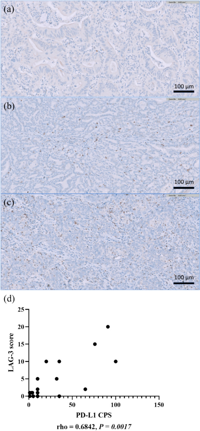
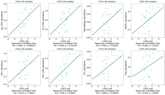
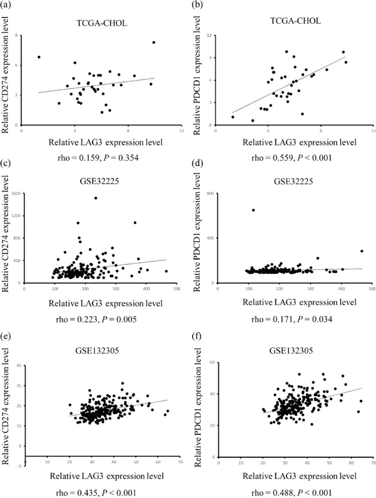
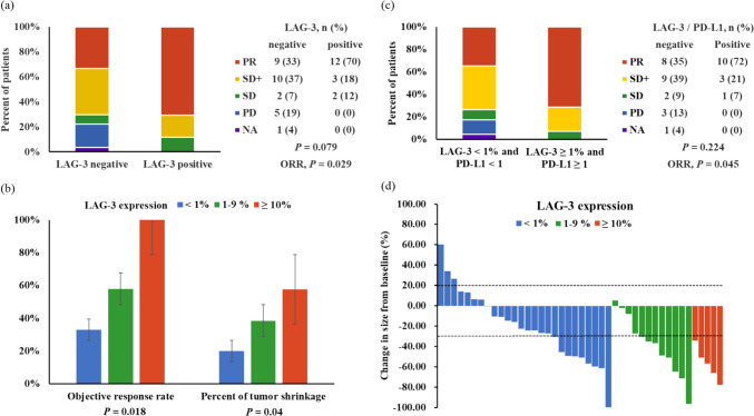
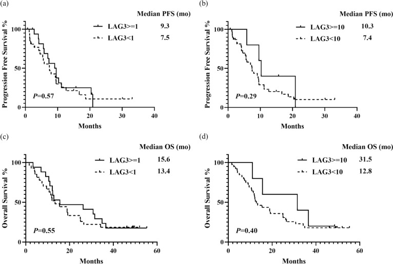
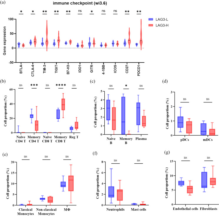
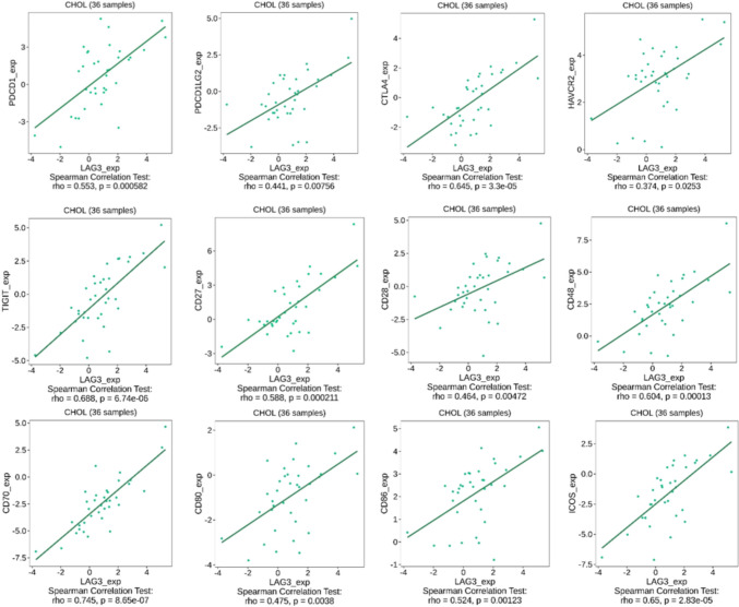
In our previous phase II T1219 trial for advanced biliary tract cancer (ABTC), the combination of nivolumab with modified gemcitabine and S-1 exhibited promising efficacy, while the programmed-death-ligand-1 (PD-L1) expression did not predict chemoimmunotherapy efficacy. Lymphocyte-activation-gene-3 (LAG-3), a negative immune checkpoint, is frequently co-expressed with PD-L1. This study assessed the predictive value of LAG-3 expression in ABTC patients who received chemoimmunotherapy. We analyzed 44 formalin-fixed ABTC samples using immunohistochemical staining for PD-L1 and LAG-3 and correlated them with the clinical efficacy of chemoimmunotherapy. Digital spatial profiling was conducted in selected regions of interest to examine immune cell infiltration and checkpoint expression in six cases. Three public BTC datasets were used for analysis: TCGA-CHOL, GSE32225, and GSE132305. LAG-3 positivity was observed in 38.6% of the ABTC samples and was significantly correlated with PD-L1 positivity (P < 0.001). The objective response rate (ORR) was significantly higher in LAG-3-positive tumors than in LAG-3-negative tumors (70.6% vs. 33.3%, P = 0.029). The LAG-3 expression level was associated with an increased ORR (33%, 58%, and 100% for LAG-3 < 1%, 1-9%, and ≥ 10%, respectively; P = 0.018) and a deeper therapeutic response (20.1%, 38.6%, and 57.6% for the same respective groups; P = 0.04). LAG-3 expression is positively correlated with the expression of numerous immune checkpoints. Enrichment of CD8 T cells was observed in LAG-3-positive BTC, indicating that LAG-3 expression may serve as a biomarker for identifying immune-inflamed tumors and predicting the therapeutic response to chemoimmunotherapy in ABTC.
在我们之前针对晚期胆管癌(ABTC)进行的II期T1219试验中,纳武单抗与改良吉西他滨和S-1联合使用显示出有前景的疗效,而程序性死亡配体1(PD-L1)表达并不能预测化疗免疫治疗的疗效。淋巴细胞激活基因3(LAG-3)是一种负性免疫检查点,经常与PD-L1共表达。本研究评估了LAG-3表达在接受化疗免疫治疗的ABTC患者中的预测价值。我们使用免疫组织化学染色对44份福尔马林固定的ABTC样本进行了PD-L1和LAG-3分析,并将其与化疗免疫治疗的临床疗效相关联。对六个病例的选定感兴趣区域进行了数字空间分析,以检查免疫细胞浸润和检查点表达。使用三个公开的BTC数据集进行分析:TCGA-CHOL、GSE32225和GSE132305。在38.6%的ABTC样本中观察到LAG-3阳性,并且与PD-L1阳性显著相关(P < 0.001)。LAG-3阳性肿瘤的客观缓解率(ORR)显著高于LAG-3阴性肿瘤(70.6%对33.3%,P = 0.029)。LAG-3表达水平与ORR增加相关(LAG-3 < 1%、1-9%和≥ 10%时分别为33%、58%和100%;P = 0.018)以及更深的治疗反应(相同分组时分别为20.1%、38.6%和57.6%;P = 0.04)。LAG-3表达与众多免疫检查点的表达呈正相关。在LAG-3阳性的BTC中观察到CD8 T细胞富集,表明LAG-3表达可能作为一种生物标志物,用于识别免疫炎症性肿瘤并预测ABTC患者对化疗免疫治疗的反应。