Okamura Kazuyuki, Sato Miyuki, Suzuki Takehiro, Nohara Keiko
Health and Environmental Risk Division, National Institute for Environmental Studies.
Environ Health Prev Med. 2024;29:74. doi: 10.1265/ehpm.24-00139.
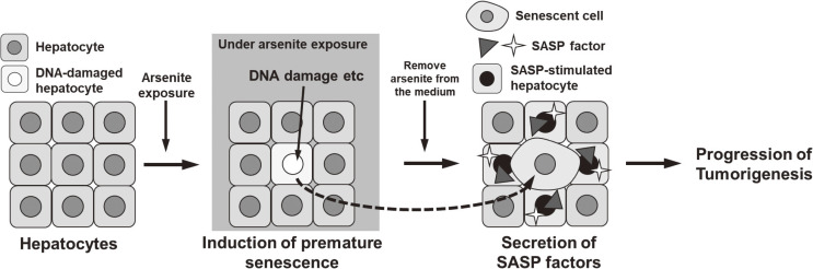
Chronic arsenite exposure has been known to induce cancer in various organs; however, the underlying mechanisms remain elusive. The characteristic feature of carcinogenesis due to arsenic exposure is that the disease develops after a prolonged latent period, even after cessation of exposure. Our previous study revealed that arsenite exposure induces premature senescence in hepatic stellate cells and suggests that the senescence-associated secretory phenotype (SASP) factors from the senescent cells promote hepatic carcinogenesis. However, arsenite exposure in the liver occurs not only in hepatic stellate cells, but also in hepatocytes. Therefore, we examined whether arsenite exposure in hepatocytes also causes premature senescence and the enhancement of SASP factors. We also assessed whether those effects remained after cessation of arsenite exposure.
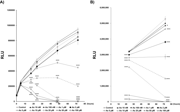
Human hepatocyte-derived cell line Huh-7 was exposed to sodium arsenite for 72 hours to determine the concentration at which cell proliferation was inhibited. In the 5 µM of exposure, various cellular senescence markers and SASP factors were analyzed and compared with unexposed cells. We also examined whether those senescence markers and SASP factors were maintained after cessation of arsenite exposure. Finally, we explored whether the increased expression of SASP factor, which was upregulated in hepatocytes by arsenic exposure in this study, is related to the prognosis of human hepatocellular carcinoma.
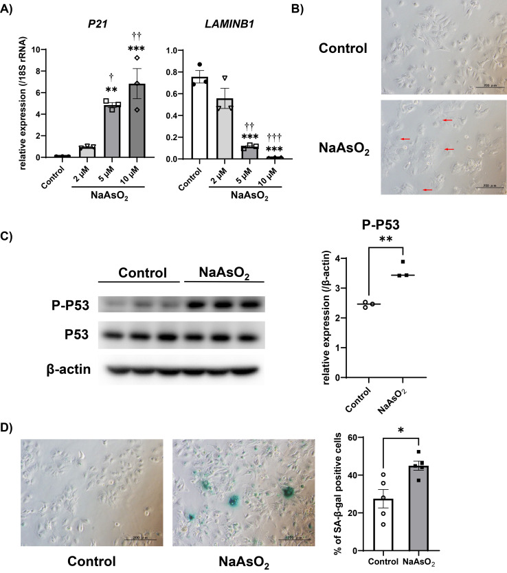
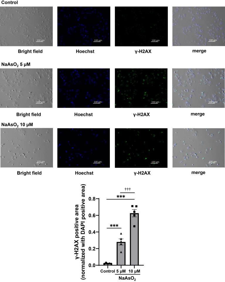
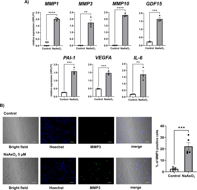
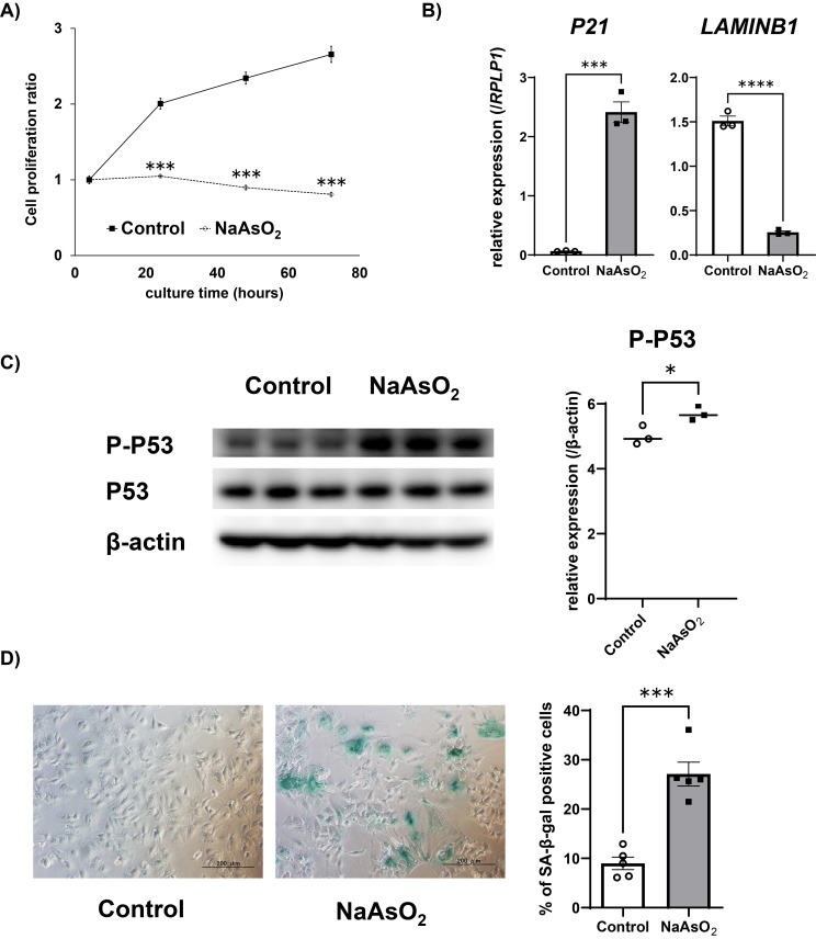
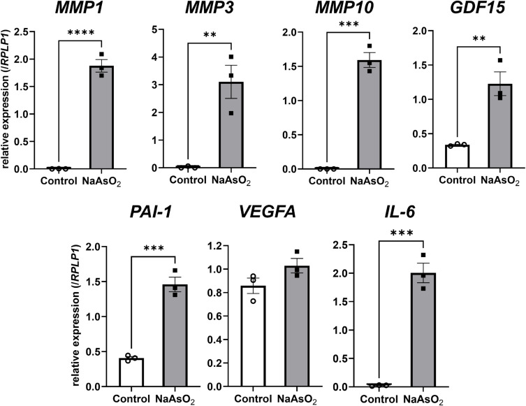
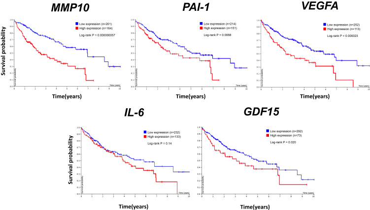
After exposure to 5 µM of sodium arsenite for 72 hours, various senescent features, such as the induction of P21 mRNA, the reduction of LAMINB1 mRNA, morphological changes, phosphorylation of P53, and the presence of SA-β-gal positive cells were observed. Those changes were maintained after cessation of arsenite exposure. In addition, mRNA levels of SASP factors (MMP1, MMP3, MMP10, GDF15, PAI-1, and IL-6) were increased after arsenite exposure, and their high expression levels were maintained after cessation of arsenite exposure. Furthermore, by analyzing the TCGA database, we found that the increased expression levels of many SASP factors negatively correlated with prognosis.
Arsenite exposure induces premature senescence in hepatocyte-derived cells and increases SASP factors that are related to hepatic tumorigenesis. Once arsenite exposure induces premature senescence, the senescent cells remain even after cessation of exposure.
长期接触亚砷酸盐已知会诱发多个器官的癌症;然而,其潜在机制仍不清楚。砷暴露致癌的特征是,即使在停止接触后,疾病也会在漫长的潜伏期后发生。我们之前的研究表明,亚砷酸盐暴露会诱导肝星状细胞过早衰老,并提示衰老细胞的衰老相关分泌表型(SASP)因子会促进肝癌发生。然而,肝脏中的亚砷酸盐暴露不仅发生在肝星状细胞中,也发生在肝细胞中。因此,我们研究了肝细胞中的亚砷酸盐暴露是否也会导致过早衰老以及SASP因子的增加。我们还评估了亚砷酸盐暴露停止后这些影响是否依然存在。
将人肝细胞系Huh-7暴露于亚砷酸钠72小时,以确定抑制细胞增殖的浓度。在5μM暴露条件下,分析各种细胞衰老标志物和SASP因子,并与未暴露细胞进行比较。我们还研究了亚砷酸盐暴露停止后这些衰老标志物和SASP因子是否依然存在。最后,我们探讨了本研究中因砷暴露而在肝细胞中上调的SASP因子表达增加是否与人类肝细胞癌的预后相关。
在暴露于5μM亚砷酸钠72小时后,观察到各种衰老特征,如P21 mRNA的诱导、LAMINB1 mRNA的减少、形态变化、P53的磷酸化以及SA-β-gal阳性细胞的存在。亚砷酸盐暴露停止后,这些变化依然存在。此外,亚砷酸盐暴露后SASP因子(MMP1、MMP3、MMP10、GDF15、PAI-1和IL-6)的mRNA水平升高,且在亚砷酸盐暴露停止后其高表达水平依然保持。此外,通过分析TCGA数据库,我们发现许多SASP因子表达水平的增加与预后呈负相关。
亚砷酸盐暴露会诱导肝细胞系细胞过早衰老,并增加与肝肿瘤发生相关的SASP因子。一旦亚砷酸盐暴露诱导过早衰老,即使在暴露停止后,衰老细胞依然存在。