Ortiz Andrés Felipe Herrera, Cuenca Nury Tatiana Rincón, Herazo Valeria Del Castillo, Jiménez Diana Marcela Oliveros, Maestre Laura García, Posada Mary Eugenia, Moreno Luz Angela
Radiology, Fundación Santa Fe de Bogotá, Bogotá, Colombia.
Radiology, Universidad El Bosque, Bogotá, Colombia.
Radiol Case Rep. 2024 Dec 12;20(3):1294-1297. doi: 10.1016/j.radcr.2024.11.040. eCollection 2025 Mar.
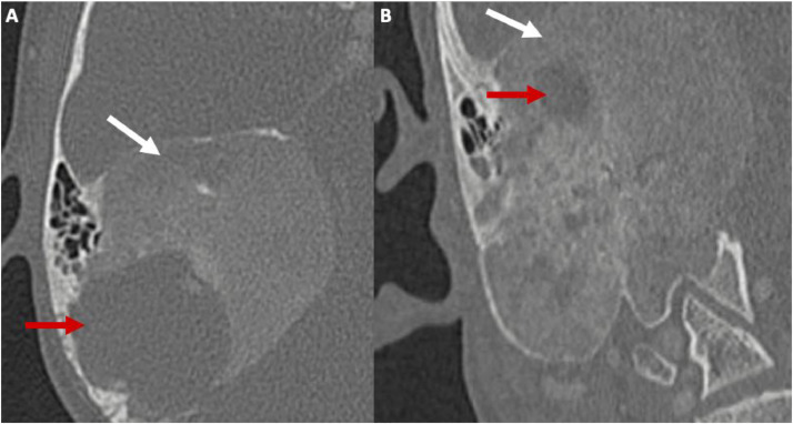
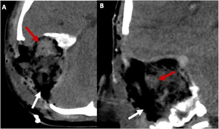
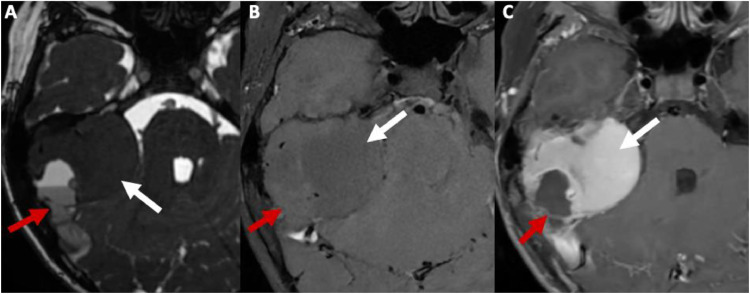
The coexistence of fibrous dysplasia and aneurysmal bone cyst in the craniofacial region is extremely rare. This article presents a unique case involving the skull base, diagnosed using computed tomography and magnetic resonance. Imaging revealed a characteristic expansile mass with a ground-glass appearance alongside cystic areas exhibiting fluid-fluid levels, indicative of fibrous dysplasia and aneurysmal bone cyst. Surgical resection was performed successfully, although significant intraoperative hemorrhage was encountered, leading to hypovolemic shock and cardiorespiratory arrest. This case underscores the essential role of imaging in accurate diagnosis and highlights the bleeding risks associated with surgical treatment of fibrous dysplasia with aneurysmal bone cyst. Preoperative embolization is suggested as a strategy to mitigate bleeding risk during surgery.
纤维发育不良与动脉瘤样骨囊肿在颅面部区域同时存在极为罕见。本文介绍了一例累及颅底的独特病例,通过计算机断层扫描和磁共振成像进行诊断。影像学检查显示有一个特征性的膨胀性肿块,呈磨玻璃样外观,同时伴有出现液-液平面的囊性区域,提示为纤维发育不良和动脉瘤样骨囊肿。尽管术中出现大量出血,导致低血容量性休克和心肺骤停,但手术切除仍成功完成。该病例强调了影像学在准确诊断中的重要作用,并突出了纤维发育不良合并动脉瘤样骨囊肿手术治疗相关的出血风险。建议术前进行栓塞,作为降低手术中出血风险的一种策略。