Pabon Maria A, Misra Amrit, Gauvreau Kimberlee, Duncan Madeline E, Conklin Ava, Economy Katherine E, Wu Fred M, Tadros Thomas, Valente Anne Marie
Division of Cardiology, Department of Medicine, Brigham and Women's Hospital, Boston, Massachusetts, USA.
Department of Cardiology, Boston Children's Hospital, Boston, Massachusetts, USA.
Ann Noninvasive Electrocardiol. 2025 Jan;30(1):e70037. doi: 10.1111/anec.70037.
Electrocardiograms (EKGs) are routinely performed in pregnant patients with pre-existing cardiovascular disease. However, in pregnant patients with congenital heart disease (CHD), EKG changes during gestation have not been explored.
We performed a retrospective study of pregnant patients with CHD enrolled in the STORCC initiative. Patients were included if they had at least two EKGs across the perinatal period and were grouped by specific conditions: atrial septal defect (ASD), tetralogy of Fallot, congenital pulmonary stenosis, coarctation of the aorta (CoA), bicuspid aortic valve (BAV), systemic right ventricle (SRV), and Fontan circulation. EKG parameters were measured in all available EKGs by two investigators, blinded to diagnosis and time of gestation.
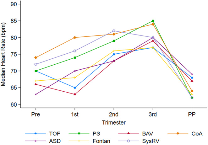
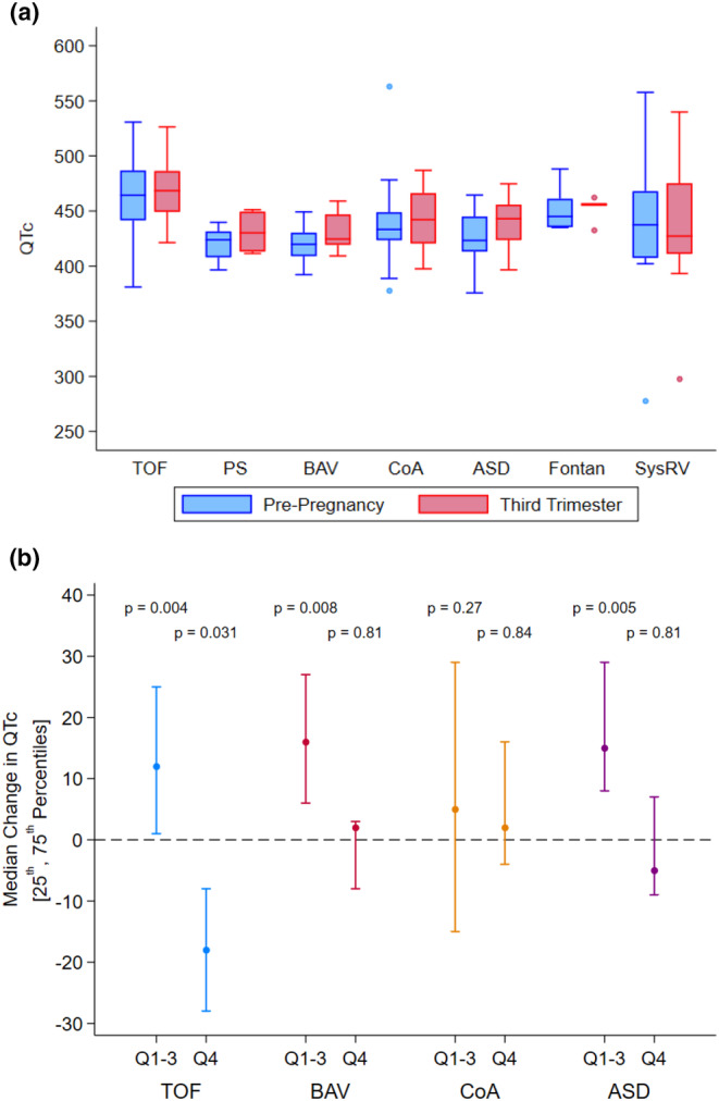
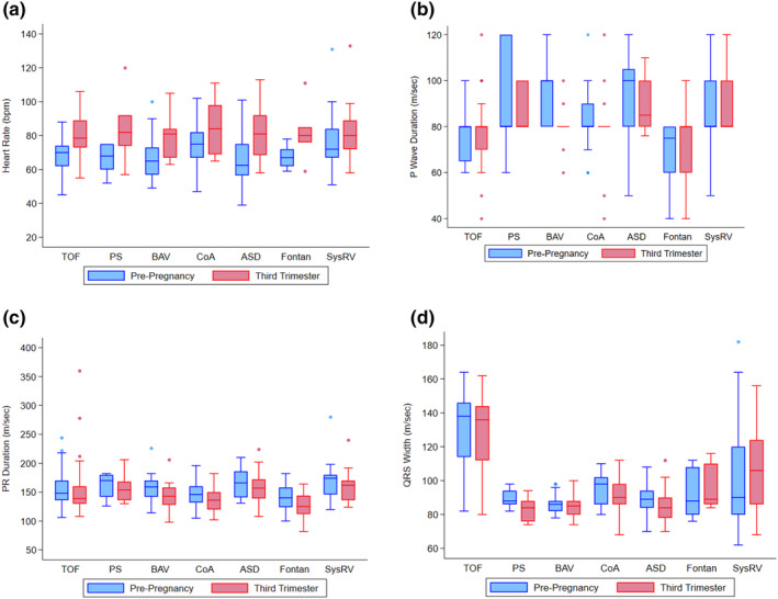
One hundred and seventy pregnant patients were included. There was a statistically significant increase in HR from pre-pregnancy to third trimester in all groups except for those with Fontan and SRV. Patients with ASD and BAV had a statistically significant increase in their QTc (ASD:13 ms, p = 0.017; BAV:7 ms, p = 0.018) during pregnancy. QRS duration was shorter (4 ms) in the third trimester for patients with ASD (p = 0.033) and CoA (p = 0.014). Despite these individual findings, EKG parameters remained within normal limits and regressed to baseline in the postpartum period.
Patients with CHD have statistically significant EKG changes throughout pregnancy, but the values remain within normal limits. Like patients without heart disease, those with CHD increase their HR during pregnancy, except individuals with SRV and Fontan, who appear to lack capacity for physiologic HR augmentation.
对于患有心血管疾病的孕妇,常规会进行心电图(EKG)检查。然而,对于患有先天性心脏病(CHD)的孕妇,孕期心电图的变化尚未得到研究。
我们对参与STORCC计划的患有CHD的孕妇进行了一项回顾性研究。纳入标准为围产期至少有两份心电图的患者,并按特定病情分组:房间隔缺损(ASD)、法洛四联症、先天性肺动脉狭窄、主动脉缩窄(CoA)、二叶式主动脉瓣(BAV)、右心室双出口(SRV)和Fontan循环。由两名对诊断和孕周不知情的研究人员测量所有可用心电图的心电图参数。
共纳入170名孕妇。除Fontan和SRV组外,所有组从孕前到孕晚期心率均有统计学意义的增加。ASD和BAV患者孕期QTc有统计学意义的增加(ASD:13毫秒,p = 0.017;BAV:7毫秒,p = 0.018)。ASD(p = 0.033)和CoA(p = 0.014)患者孕晚期QRS时限缩短(4毫秒)。尽管有这些个体差异,但心电图参数仍在正常范围内,并在产后恢复至基线水平。
患有CHD的患者在整个孕期心电图有统计学意义的变化,但数值仍在正常范围内。与无心脏病的患者一样,患有CHD的患者孕期心率会增加,但SRV和Fontan患者似乎缺乏生理性心率增加的能力。