Cappelleri Andrea, Brambilla Eleonora, Chiti Lavinia E, Trapletti Alessia, Bianchi Gaia B M, Di Giancamillo Mauro, Grieco Valeria, Giudice Chiara
Department of Veterinary Medicine and Animal Sciences, University of Milan, 26900 Lodi, Italy.
Clinic for Small Animal Surgery, Department for Small Animals, Vetsuisse Faculty, University of Zurich, 8057 Zurich, Switzerland.
Animals (Basel). 2024 Dec 12;14(24):3588. doi: 10.3390/ani14243588.
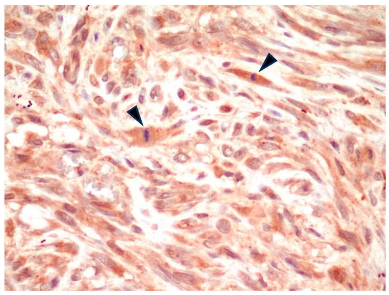
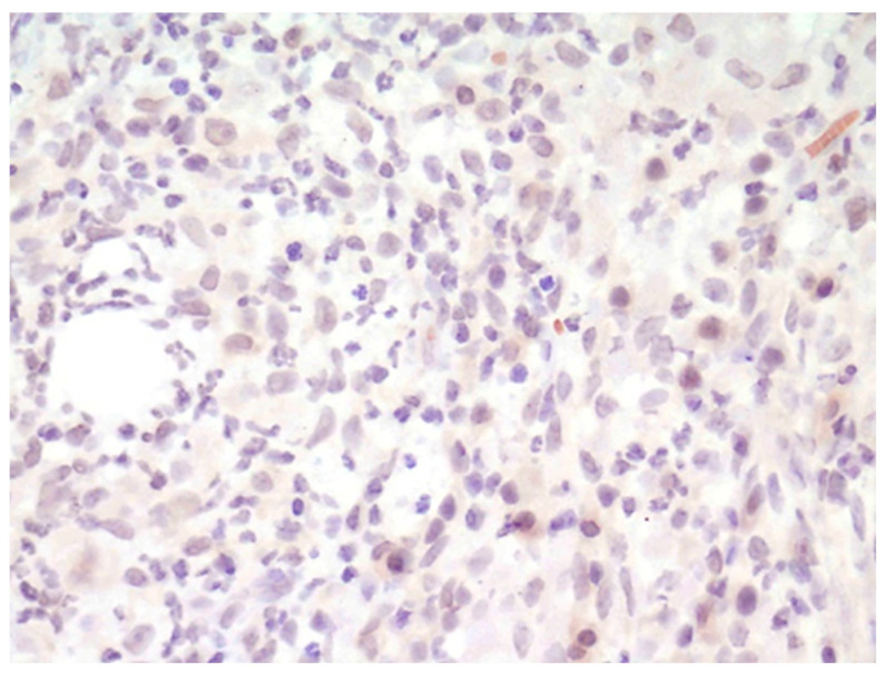
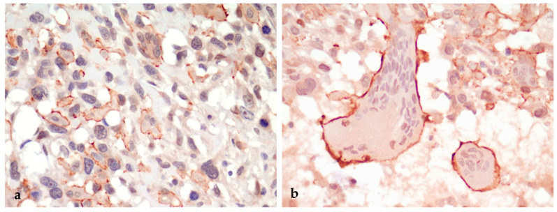
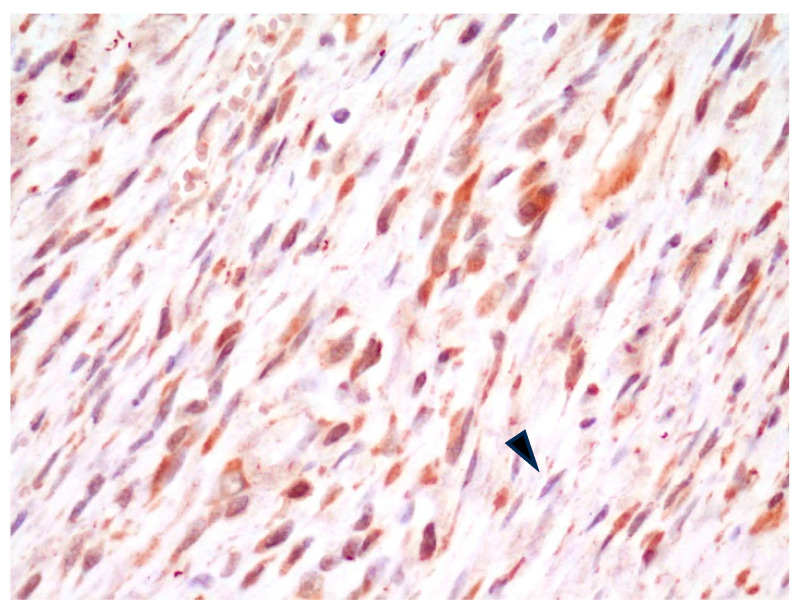
Feline injection-site sarcomas (FISSs) are malignant skin tumors of mesenchymal origin arising at local post-vaccination (or injection) sites. In recent years, a fluorescence imaging technique based on probes targeting αβ integrin has been effectively applied for the surgical complete resection of the tumor. In our study, we investigated the utility of a commercially available anti-α integrin polyclonal antibody for the histopathological evaluation of FISS's surgical excision margins. We collected 10 formalin-fixed paraffin-embedded (FFPE) feline excisional biopsies with a histopathological diagnosis of FISS (7 fibrosarcomas and 3 pleomorphic sarcomas) and wide margin tissue, along with one subcutaneous injection-site granuloma and 6 osteosarcomas. Samples were processed for histology, and slides were stained for IHC with the anti-α integrin antibody. Immunostained slides were evaluated for the cellular localization and intensity of the staining in different neoplastic and non-neoplastic cell populations. Neoplastic and non-neoplastic spindle cells had cytoplasmic positivity in all fibrosarcomas. Inflammatory cells, including macrophages of the injection-site granuloma, were negative. Multinucleated giant cells in the pleomorphic sarcomas had an intense membranous positivity. Although the anti-α integrin antibody was ineffective for the histopathological evaluation of surgical excision margins, the membranous localization of α integrin in multinucleated giant cells of pleomorphic sarcomas suggests that it plays a role in the oncogenesis of this FISS variant.
猫注射部位肉瘤(FISSs)是一种间充质来源的恶性皮肤肿瘤,发生于局部疫苗接种(或注射)部位。近年来,一种基于靶向αβ整合素探针的荧光成像技术已被有效地应用于肿瘤的手术完全切除。在我们的研究中,我们研究了一种市售的抗α整合素多克隆抗体在FISS手术切除边缘组织病理学评估中的效用。我们收集了10例经组织病理学诊断为FISS的福尔马林固定石蜡包埋(FFPE)猫切除活检标本(7例纤维肉瘤和3例多形性肉瘤)及宽切缘组织,以及1例皮下注射部位肉芽肿和6例骨肉瘤。对样本进行组织学处理,并用抗α整合素抗体对切片进行免疫组化染色。对免疫染色切片评估不同肿瘤性和非肿瘤性细胞群体中染色的细胞定位和强度。在所有纤维肉瘤中,肿瘤性和非肿瘤性梭形细胞均呈细胞质阳性。包括注射部位肉芽肿中的巨噬细胞在内的炎症细胞呈阴性。多形性肉瘤中的多核巨细胞呈强烈的膜阳性。尽管抗α整合素抗体对手术切除边缘的组织病理学评估无效,但α整合素在多形性肉瘤多核巨细胞中的膜定位表明它在这种FISS变体的肿瘤发生中起作用。