• 文献检索
  • 文档翻译
  • 深度研究
  • 学术资讯
  • Suppr Zotero 插件Zotero 插件
  • 邀请有礼
  • 套餐&价格
  • 历史记录
应用&插件
Suppr Zotero 插件Zotero 插件浏览器插件Mac 客户端Windows 客户端微信小程序
定价
高级版会员购买积分包购买API积分包
服务
文献检索文档翻译深度研究API 文档MCP 服务
关于我们
关于 Suppr公司介绍联系我们用户协议隐私条款
关注我们

Suppr 超能文献

核心技术专利:CN118964589B侵权必究
粤ICP备2023148730 号-1Suppr @ 2026

文献检索

告别复杂PubMed语法,用中文像聊天一样搜索,搜遍4000万医学文献。AI智能推荐,让科研检索更轻松。

立即免费搜索

文件翻译

保留排版,准确专业,支持PDF/Word/PPT等文件格式,支持 12+语言互译。

免费翻译文档

深度研究

AI帮你快速写综述,25分钟生成高质量综述,智能提取关键信息,辅助科研写作。

立即免费体验

使用深度学习模型对非增强胸部CT进行心脏腔室容积和心肌质量的全自动评估:与心脏磁共振成像的对比验证

Fully Automated Assessment of Cardiac Chamber Volumes and Myocardial Mass on Non-Contrast Chest CT with a Deep Learning Model: Validation Against Cardiac MR.

作者信息

Schmitt Ramona, Schlett Christopher L, Sperl Jonathan I, Rapaka Saikiran, Jacob Athira J, Hein Manuel, Hagar Muhammad Taha, Ruile Philipp, Westermann Dirk, Soschynski Martin, Bamberg Fabian, Schuppert Christopher

机构信息

Department of Cardiology and Angiology, University Heart Center Freiburg-Bad Krozingen, Medical Center-University of Freiburg, Faculty of Medicine, University of Freiburg, Südring 15, 79189 Bad Krozingen, Germany.

Department of Diagnostic and Interventional Radiology, Medical Center-University of Freiburg, Faculty of Medicine, University of Freiburg, Hugstetter Str. 55, 79106 Freiburg im Breisgau, Germany.

出版信息

Diagnostics (Basel). 2024 Dec 21;14(24):2884. doi: 10.3390/diagnostics14242884.

DOI:10.3390/diagnostics14242884
PMID:39767245
原文链接:https://pmc.ncbi.nlm.nih.gov/articles/PMC11675647/
Abstract

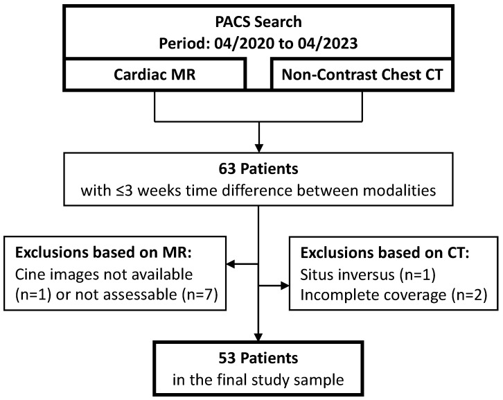
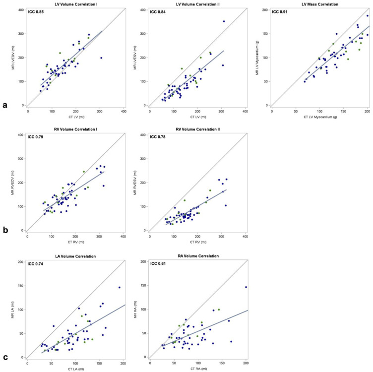
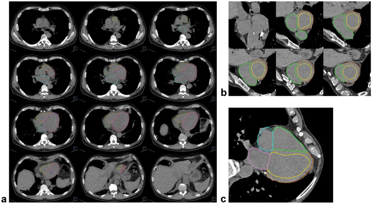
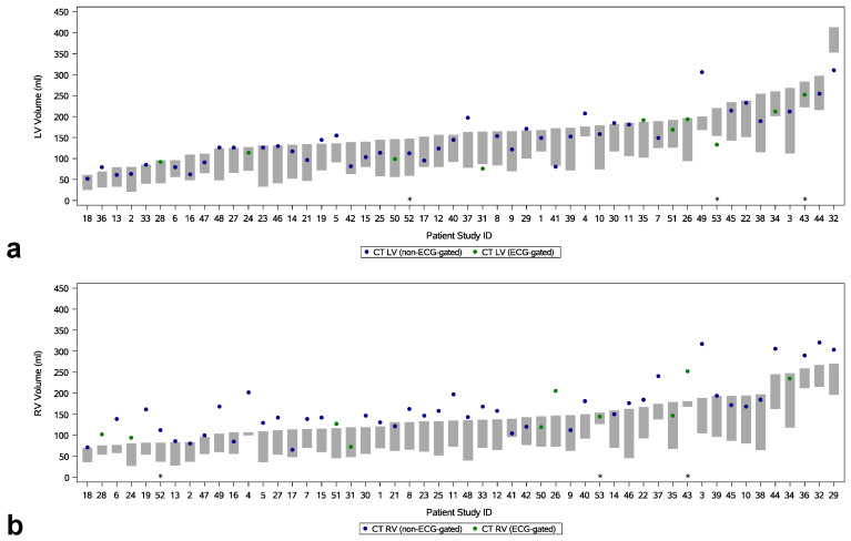
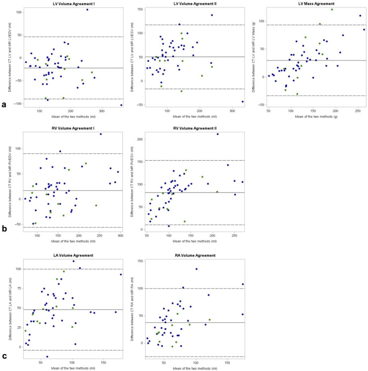
: To validate the automated quantification of cardiac chamber volumes and myocardial mass on non-contrast chest CT using cardiac MR (CMR) as a reference. : We retrospectively included 53 consecutive patients who received non-contrast chest CT and CMR within three weeks. A deep learning model created cardiac segmentations on axial soft-tissue reconstructions from CT, covering all four cardiac chambers and the left ventricular myocardium. Segmentations on CMR cine short-axis and long-axis images served as a reference. Standard estimates of diagnostic accuracy were calculated for ventricular volumes at end-diastole and end-systole (LVEDV, LVESV, RVEDV, RVESV), left ventricular mass (LVM), and atrial volumes (LA, RA) at ventricular end-diastole. A qualitative assessment noted segmentation issues. : The deep learning model generated CT measurements for 52 of the 53 patients (98%). Based on CMR measurements, the average LVEDV was 166 ± 64 mL, RVEDV was 144 ± 51 mL, and LVM was 115 ± 39 g. The CT measurements correlated well with CMR measurements for LVEDV, LVESV, and LVM (ICC = 0.85, ICC = 0.84, and ICC = 0.91; all < 0.001) and RVEDV and RVESV (ICC = 0.79 and ICC= 0.78; both < 0.001), and moderately well with LA and RA (ICC = 0.74 and ICC = 0.61; both < 0.001). Absolute agreements likewise favored LVEDV, LVM, and RVEDV. ECG-gating did not relevantly influence the results. The CT results correctly identified 7/15 LV and 1/1 RV as dilated (one and six false positives, respectively). Major qualitative issues were found in three cases (6%). : Automated cardiac chamber volume and myocardial mass quantification on non-contrast chest CT produced viable measurements in this retrospective sample. : An automated cardiac assessment on non-contrast chest CT provides quantitative morphological data on the heart, enabling a preliminary organ evaluation that aids in incidentally identifying at-risk patients who may benefit from a more targeted diagnostic workup.

摘要

目的

以心脏磁共振成像(CMR)为参考,验证非增强胸部CT对心腔容积和心肌质量的自动定量分析。

方法

我们回顾性纳入了53例在三周内接受非增强胸部CT和CMR检查的连续患者。一个深度学习模型在CT的轴向软组织重建图像上创建心脏分割,覆盖所有四个心腔和左心室心肌。CMR电影短轴和长轴图像上的分割作为参考。计算舒张末期和收缩末期心室容积(左心室舒张末期容积、左心室收缩末期容积、右心室舒张末期容积、右心室收缩末期容积)、左心室质量以及舒张末期心房容积(左心房、右心房)的诊断准确性标准估计值。进行定性评估以发现分割问题。

结果

深度学习模型为53例患者中的52例(98%)生成了CT测量值。基于CMR测量值,平均左心室舒张末期容积为166±64 mL,右心室舒张末期容积为144±51 mL,左心室质量为115±39 g。CT测量值与CMR测量值在左心室舒张末期容积、左心室收缩末期容积和左心室质量方面相关性良好(组内相关系数ICC = 0.85、ICC = 0.84和ICC = 0.91;均P < 0.001),在右心室舒张末期容积和右心室收缩末期容积方面也相关性良好(ICC = 0.79和ICC = 0.78;均P < 0.001),与左心房和右心房的相关性中等(ICC = 0.74和ICC = 0.61;均P < 0.001)。绝对一致性同样有利于左心室舒张末期容积、左心室质量和右心室舒张末期容积。心电图门控对结果无显著影响。CT结果正确识别出7/15的左心室和1/1的右心室扩张(分别有1例和6例假阳性)。在3例(6%)中发现了主要的定性问题。

结论

在这个回顾性样本中,非增强胸部CT上心腔容积和心肌质量的自动定量分析产生了可行的测量结果。

非增强胸部CT上的自动心脏评估提供了心脏的定量形态学数据,有助于进行初步的器官评估,从而有助于偶然识别可能从更有针对性的诊断检查中受益的高危患者。

https://cdn.ncbi.nlm.nih.gov/pmc/blobs/2f67/11675647/fad3b3613021/diagnostics-14-02884-g005.jpg
https://cdn.ncbi.nlm.nih.gov/pmc/blobs/2f67/11675647/f28447858558/diagnostics-14-02884-g001.jpg
https://cdn.ncbi.nlm.nih.gov/pmc/blobs/2f67/11675647/589c7c41ea7a/diagnostics-14-02884-g002.jpg
https://cdn.ncbi.nlm.nih.gov/pmc/blobs/2f67/11675647/9001bc6fa87d/diagnostics-14-02884-g003.jpg
https://cdn.ncbi.nlm.nih.gov/pmc/blobs/2f67/11675647/b5213e752dbd/diagnostics-14-02884-g004.jpg
https://cdn.ncbi.nlm.nih.gov/pmc/blobs/2f67/11675647/fad3b3613021/diagnostics-14-02884-g005.jpg

相似文献

1
Fully Automated Assessment of Cardiac Chamber Volumes and Myocardial Mass on Non-Contrast Chest CT with a Deep Learning Model: Validation Against Cardiac MR.使用深度学习模型对非增强胸部CT进行心脏腔室容积和心肌质量的全自动评估:与心脏磁共振成像的对比验证
Diagnostics (Basel). 2024 Dec 21;14(24):2884. doi: 10.3390/diagnostics14242884.
2
Automated cardiovascular magnetic resonance image analysis with fully convolutional networks.基于全卷积网络的自动化心血管磁共振图像分析。
J Cardiovasc Magn Reson. 2018 Sep 14;20(1):65. doi: 10.1186/s12968-018-0471-x.
3
Fully automated quantification of cardiac chamber and function assessment in 2-D echocardiography: clinical feasibility of deep learning-based algorithms.二维超声心动图中心腔和功能评估的全自动化定量:基于深度学习算法的临床可行性
Int J Cardiovasc Imaging. 2022 May;38(5):1047-1059. doi: 10.1007/s10554-021-02482-y. Epub 2022 Feb 13.
4
Automated left and right ventricular chamber segmentation in cardiac magnetic resonance images using dense fully convolutional neural network.使用密集全卷积神经网络对心脏磁共振图像进行自动左右心室腔分割
Comput Methods Programs Biomed. 2021 Jun;204:106059. doi: 10.1016/j.cmpb.2021.106059. Epub 2021 Mar 21.
5
Validating real-time three-dimensional echocardiography against cardiac magnetic resonance, for the determination of ventricular mass, volume and ejection fraction: a meta-analysis.实时三维超声心动图与心脏磁共振对比评估心室质量、容积和射血分数的验证:荟萃分析。
Clin Res Cardiol. 2024 Mar;113(3):367-392. doi: 10.1007/s00392-023-02204-5. Epub 2023 Apr 20.
6
Multi-detector row cardiac computed tomography accurately quantifies right and left ventricular size and function compared with cardiac magnetic resonance.与心脏磁共振成像相比,多排探测器心脏计算机断层扫描能准确量化右心室和左心室的大小及功能。
Am Heart J. 2006 Mar;151(3):736-44. doi: 10.1016/j.ahj.2005.04.029.
7
Feasibility of New Transthoracic Three-Dimensional Echocardiographic Automated Software for Left Heart Chamber Quantification in Children.新型经胸三维超声心动图自动软件用于儿童左心腔定量的可行性。
J Am Soc Echocardiogr. 2019 Jan;32(1):121-134.e1. doi: 10.1016/j.echo.2018.08.001. Epub 2018 Sep 18.
8
Development and validation of AI-derived segmentation of four-chamber cine cardiac magnetic resonance.四腔心电影心脏磁共振人工智能分割的研发与验证。
Eur Radiol Exp. 2024 Jul 12;8(1):77. doi: 10.1186/s41747-024-00477-7.
9
Evaluation of deep learning estimation of whole heart anatomy from automated cardiovascular magnetic resonance short- and long-axis analyses in UK Biobank.基于英国生物库自动心血管磁共振短轴和长轴分析的深度学习估计全心解剖结构的评估。
Eur Heart J Cardiovasc Imaging. 2024 Sep 30;25(10):1374-1383. doi: 10.1093/ehjci/jeae123.
10
Cardiac chamber volumes, function, and mass as determined by 64-multidetector row computed tomography: mean values among healthy adults free of hypertension and obesity.64排多层螺旋计算机断层扫描测定的心脏腔室容积、功能及质量:无高血压和肥胖的健康成年人的平均值
JACC Cardiovasc Imaging. 2008 Nov;1(6):782-6. doi: 10.1016/j.jcmg.2008.04.015.

引用本文的文献

1
[New innovations in cross-sectional imaging diagnostics of the aorta].[主动脉横断面成像诊断的新进展]
Radiologie (Heidelb). 2025 Mar;65(3):201-214. doi: 10.1007/s00117-025-01424-2.

本文引用的文献

1
Artificial intelligence applied to coronary artery calcium scans (AI-CAC) significantly improves cardiovascular events prediction.应用于冠状动脉钙化扫描的人工智能(AI-CAC)显著改善心血管事件预测。
NPJ Digit Med. 2024 Nov 5;7(1):309. doi: 10.1038/s41746-024-01308-0.
2
AI-Derived Left Ventricular Mass From Noncontrast Cardiac CT: Correlation With Contrast CT Angiography and CMR.基于非增强心脏CT的人工智能衍生左心室质量:与增强CT血管造影和心脏磁共振成像的相关性
JACC Adv. 2024 Sep 12;3(10):101249. doi: 10.1016/j.jacadv.2024.101249. eCollection 2024 Oct.
3
Predicting mortality from AI cardiac volumes mass and coronary calcium on chest computed tomography.
基于胸部 CT 的人工智能心腔容积质量和冠状动脉钙分数预测死亡率。
Nat Commun. 2024 Mar 29;15(1):2747. doi: 10.1038/s41467-024-46977-3.
4
Deep learning-based automatic segmentation of cardiac substructures for lung cancers.基于深度学习的肺癌心脏亚结构自动分割
Radiother Oncol. 2024 Feb;191:110061. doi: 10.1016/j.radonc.2023.110061. Epub 2023 Dec 19.
5
AI-based, automated chamber volumetry from gated, non-contrast CT.基于人工智能的门控非对比 CT 全自动心腔容积测量。
J Cardiovasc Comput Tomogr. 2023 Sep-Oct;17(5):336-340. doi: 10.1016/j.jcct.2023.08.001. Epub 2023 Aug 21.
6
Comparison of Biplane Area-Length Method and 3D Volume Quantification by Using Cardiac MRI for Assessment of Left Atrial Volume in Atrial Fibrillation.使用心脏磁共振成像比较双平面面积-长度法和三维容积定量法评估心房颤动患者左心房容积
Radiol Cardiothorac Imaging. 2023 Apr 13;5(2):e220133. doi: 10.1148/ryct.220133. eCollection 2023 Apr.
7
Fully automatic cardiac four chamber and great vessel segmentation on CT pulmonary angiography using deep learning.基于深度学习的CT肺血管造影中全自动心脏四腔心及大血管分割
Front Cardiovasc Med. 2022 Sep 26;9:983859. doi: 10.3389/fcvm.2022.983859. eCollection 2022.
8
2022 ESC Guidelines on cardio-oncology developed in collaboration with the European Hematology Association (EHA), the European Society for Therapeutic Radiology and Oncology (ESTRO) and the International Cardio-Oncology Society (IC-OS).2022年欧洲心脏病学会(ESC)与欧洲血液学协会(EHA)、欧洲治疗放射学与肿瘤学协会(ESTRO)以及国际心脏肿瘤学会(IC-OS)合作制定的心脏肿瘤学指南。
Eur Heart J. 2022 Nov 1;43(41):4229-4361. doi: 10.1093/eurheartj/ehac244.
9
Diagnostic accuracy of automated 3D volumetry of cardiac chambers by CT pulmonary angiography for identification of pulmonary hypertension due to left heart disease.CT 肺动脉造影自动 3D 心腔容积测量对左心疾病所致肺动脉高压的诊断准确性。
Eur Radiol. 2022 Aug;32(8):5222-5232. doi: 10.1007/s00330-022-08663-0. Epub 2022 Mar 10.
10
Imaging assessment of the right atrium: anatomy and function.右心房的影像学评估:解剖与功能。
Eur Heart J Cardiovasc Imaging. 2022 Jun 21;23(7):867-884. doi: 10.1093/ehjci/jeac011.