Somnin Chutintorn, Chamieh Joseph, Leclercq Laurent, Medina Christelle, Rousseaux Olivier, Cottet Hervé
IBMM, University of Montpellier, CNRS, ENSCM, 34095 Montpellier, France.
GUERBET, Research and Innovation, 16 rue Jean Chaptal, 93600 Aulnay Sous Bois, France.
Pharmaceuticals (Basel). 2024 Dec 5;17(12):1633. doi: 10.3390/ph17121633.
Gadolinium-based contrast agents (GBCA) are widely used in magnetic resonance imaging (MRI) to enhance image contrast by interacting with water molecules, thus improving diagnostic capabilities. However, understanding the residual accumulation of GBCA in tissues after administration remains an area of active research. This highlights the need for advanced analytical techniques capable of investigating interactions between GBCAs and biopolymers, such as type I collagen, which are abundant in the body.
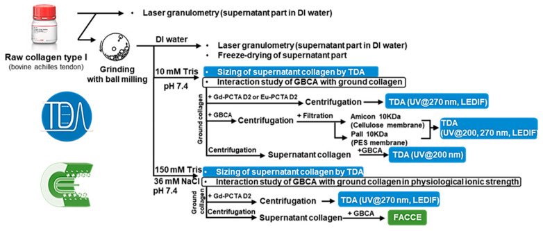
This study explores the interactions of neutral and charged GBCAs with type I collagen under physiological pH conditions (pH 7.4) using Taylor dispersion analysis (TDA) and frontal analysis continuous capillary electrophoresis (FACCE).
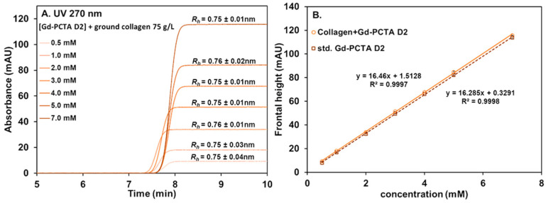
Collagen from bovine achilles tendon was ground using a vibratory ball mill to achieve a more uniform particle size and increased surface area. Laser granulometry was employed to characterize the size distributions of both raw and ground collagen suspensions in water. TDA was used to assess the hydrodynamic radius () of the soluble collagen fraction present in the supernatant.
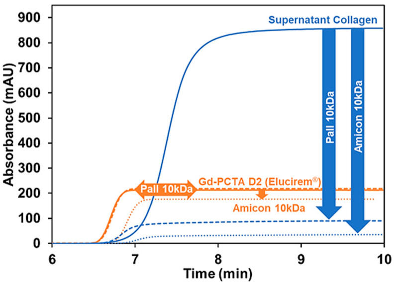
From the TDA and FACCE results, it was shown that there were no significant interactions between the tested GBCAs and either the ground collagen or its soluble fraction at pH 7.4. Interestingly, we also observed that collagen interacts with filtration membranes, indicating that careful selection of membrane material, or the absence of filtration in the experimental protocol, is essential in interaction studies involving collagen.
These findings bring valuable insights into the behavior of GBCAs in biological systems with potential implications for clinical applications.
基于钆的造影剂(GBCA)广泛应用于磁共振成像(MRI),通过与水分子相互作用来增强图像对比度,从而提高诊断能力。然而,了解给药后GBCA在组织中的残留积累仍是一个活跃的研究领域。这凸显了对先进分析技术的需求,这些技术能够研究GBCA与生物聚合物(如人体中丰富的I型胶原蛋白)之间的相互作用。
本研究使用泰勒分散分析(TDA)和前沿分析连续毛细管电泳(FACCE),探讨中性和带电GBCA在生理pH条件(pH 7.4)下与I型胶原蛋白的相互作用。
使用振动球磨机研磨牛跟腱中的胶原蛋白,以获得更均匀的粒径和更大的表面积。采用激光粒度分析来表征水中未处理和研磨后的胶原蛋白悬浮液的粒径分布。TDA用于评估上清液中可溶性胶原蛋白部分的流体动力学半径()。
从TDA和FACCE结果可知,在pH 7.4时,测试的GBCA与研磨后的胶原蛋白或其可溶部分之间没有显著相互作用。有趣的是,我们还观察到胶原蛋白与滤膜相互作用,这表明在涉及胶原蛋白的相互作用研究中,仔细选择膜材料或在实验方案中不进行过滤至关重要。
这些发现为GBCA在生物系统中的行为提供了有价值的见解,对临床应用具有潜在意义。