Silina Ekaterina V, Manturova Natalia E, Chuvilina Elena L, Gasanov Akhmedali A, Andreeva Olga I, Pugachevskii Maksim A, Kochura Aleksey V, Kryukov Alexey A, Suzdaltseva Yulia G, Stupin Victor A
Institute of Digital Biodesign and Modeling of Living Systems, I.M. Sechenov First Moscow State Medical University (Sechenov University), 119991 Moscow, Russia.
Department of Hospital Surgery, Department of Plastic and Reconstructive Surgery, Cosmetology and Cell Technology, Pirogov Russian National Research Medical University (RNRMU), 117997 Moscow, Russia.
Pharmaceutics. 2024 Dec 23;16(12):1627. doi: 10.3390/pharmaceutics16121627.
BACKGROUND/OBJECTIVES: The aim was to study the possibilities of biomedical application of gadolinium oxide nanoparticles (GdO NPs) synthesized under industrial conditions, and evaluate their physicochemical properties, redox activity, biological activity, and safety using different human cell lines.
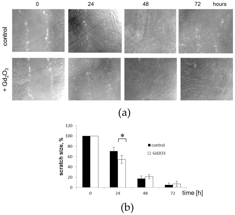
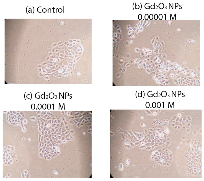
The powder of GdO NPs was obtained by a process of thermal decomposition of gadolinium carbonate precipitated from nitrate solution, and was studied using transmission electron microscopy (TEM), X-ray diffraction (XRD), Raman spectroscopy, mass spectrometry, and scanning electron microscopy (SEM) with energy dispersive X-ray analyzer (EDX). The redox activity of different concentrations of GdO NPs was studied by the optical spectroscopy (OS) method in the photochemical degradation process of methylene blue dye upon irradiation with an optical source. Biological activity was studied on different human cell lines (keratinocytes, fibroblasts, mesenchymal stem cells (MSCs)) with evaluation of the effect of a wide range of GdO NP concentrations on metabolic and proliferative cellular activity (MTT test, direct cell counting, dead cell assessment, and visual assessment of cytoarchitectonics). The test of migration activity assessment on a model wound was performed on MSC culture.
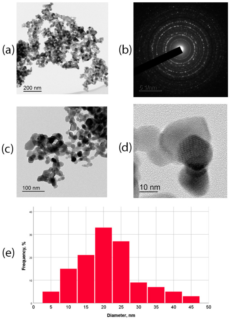
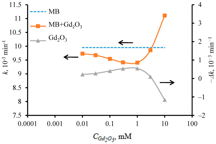
According to TEM data, the size of the NPs was in the range of 2-43 nm, with an average of 20 nm. XRD analysis revealed that the f GdO nanoparticles had a cubic structure (C-form) of GdO (Ia3)¯ with lattice parameter a = 10.79(9) Å. Raman spectroscopy showed that the f GdO nanoparticles had a high degree of crystallinity. By investigating the photooxidative degradation of methylene blue dye in the presence of f GdO NPs under red light irradiation, it was found that f GdO nanoparticles showed weak antioxidant activity, which depended on the particle content in the solution. At a concentration of 10 M, the highest antioxidant activity of f GdO nanoparticles was observed when the reaction rate constant of dye photodegradation decreased by 5.5% to 9.4 × 10 min. When the concentration of f GdO NPs in solution was increased to 10 M upon irradiation with a red light source, their antioxidant activity changed to pro-oxidant activity, accompanied by a 15% increase in the reaction rate of methylene blue degradation. Studies on cell lines showed a high level of safety and regenerative potential of GdO NPs, which stimulated fibroblast metabolism at a concentration of 10 M (27% enhancement), stimulated keratinocyte metabolism at concentrations of 10 M-10 M, and enhanced keratinocyte proliferation by an average of 35% at concentrations of 10 M. Furthermore, it accelerated the migration of MSCs, enhancing their proliferation, and promoting the healing of the model wound.
The results of the study demonstrated the safety and regenerative potential of redox-active GdO NPs towards different cell lines. This may be the basis for further research to develop nanomaterials based on GdO NPs for skin wound healing and in regenerative medicine generally.
背景/目的:本研究旨在探讨在工业条件下合成的氧化钆纳米颗粒(GdO NPs)在生物医学领域的应用可能性,并使用不同的人类细胞系评估其物理化学性质、氧化还原活性、生物活性和安全性。
通过热分解从硝酸盐溶液中沉淀出的碳酸钆来制备GdO NPs粉末,并使用透射电子显微镜(TEM)、X射线衍射(XRD)、拉曼光谱、质谱以及配备能量色散X射线分析仪(EDX)的扫描电子显微镜(SEM)对其进行研究。采用光谱法(OS)研究不同浓度的GdO NPs在光源照射下对亚甲基蓝染料光化学降解过程中的氧化还原活性。在不同的人类细胞系(角质形成细胞、成纤维细胞、间充质干细胞(MSCs))上研究生物活性,评估不同浓度范围的GdO NPs对细胞代谢和增殖活性的影响(MTT试验、直接细胞计数、死细胞评估以及细胞结构的视觉评估)。在MSC培养物上进行模型伤口迁移活性评估试验。
根据TEM数据,纳米颗粒的尺寸在2 - 43nm范围内,平均为20nm。XRD分析表明,f GdO纳米颗粒具有GdO(Ia3)¯的立方结构(C型),晶格参数a = 10.79(9) Å。拉曼光谱显示f GdO纳米颗粒具有高度结晶性。通过研究在红光照射下f GdO NPs存在时亚甲基蓝染料的光氧化降解,发现f GdO纳米颗粒表现出较弱抗氧化活性,这取决于溶液中颗粒的含量。在浓度为10 M时,当染料光降解反应速率常数降至9.4×10 min且降低5.5%时,观察到f GdO纳米颗粒的最高抗氧化活性。当在红光照射下溶液中f GdO NPs的浓度增加到10 M时,其抗氧化活性转变为促氧化活性,同时亚甲基蓝降解反应速率增加15%。对细胞系的研究表明,GdO NPs具有高度的安全性和再生潜力,在浓度为10 M时刺激成纤维细胞代谢(增强27%),在浓度为10 M - 10 M时刺激角质形成细胞代谢,在浓度为10 M时平均增强角质形成细胞增殖35%。此外,它加速了MSCs的迁移,增强了其增殖,并促进了模型伤口的愈合。
研究结果证明了具有氧化还原活性的GdO NPs对不同细胞系的安全性和再生潜力。这可能是进一步开展基于GdO NPs的纳米材料用于皮肤伤口愈合及一般再生医学研究的基础。