• 文献检索
  • 文档翻译
  • 深度研究
  • 学术资讯
  • Suppr Zotero 插件Zotero 插件
  • 邀请有礼
  • 套餐&价格
  • 历史记录
应用&插件
Suppr Zotero 插件Zotero 插件浏览器插件Mac 客户端Windows 客户端微信小程序
定价
高级版会员购买积分包购买API积分包
服务
文献检索文档翻译深度研究API 文档MCP 服务
关于我们
关于 Suppr公司介绍联系我们用户协议隐私条款
关注我们

Suppr 超能文献

核心技术专利:CN118964589B侵权必究
粤ICP备2023148730 号-1Suppr @ 2026

文献检索

告别复杂PubMed语法,用中文像聊天一样搜索,搜遍4000万医学文献。AI智能推荐,让科研检索更轻松。

立即免费搜索

文件翻译

保留排版,准确专业,支持PDF/Word/PPT等文件格式,支持 12+语言互译。

免费翻译文档

深度研究

AI帮你快速写综述,25分钟生成高质量综述,智能提取关键信息,辅助科研写作。

立即免费体验

超声超分辨率成像用于前列腺病变微血管的无创评估。

Ultrasound super-resolution imaging for non-invasive assessment of microvessel in prostate lesion.

作者信息

Huang Xin, Ye Huarong, Hu Yugang, Lei Yumeng, Tian Yi, Huang Xingyue, Zhang Jun, Zhang Yao, Gui Bin, Liu Qianhui, Zhang Ge, Deng Qing

机构信息

Department of Ultrasound Imaging, Renmin Hospital of Wuhan University, Wuhan, 430060, China.

Department of Medical Ultrasound, China Resources & Wisco General Hospital, Wuhan University of Science and Technology, Wuhan, 430080, China.

出版信息

Cancer Imaging. 2025 Jan 7;25(1):1. doi: 10.1186/s40644-024-00819-z.

DOI:10.1186/s40644-024-00819-z
PMID:39773358
原文链接:https://pmc.ncbi.nlm.nih.gov/articles/PMC11706184/
Abstract

BACKGROUND

Prostate cancer (PCa) is the leading cause of cancer-related morbidity and mortality in men worldwide. An early and accurate diagnosis is crucial for effective treatment and prognosis. Traditional invasive procedures such as image-guided prostate biopsy often cause discomfort and complications, deterring some patients from undergoing these necessary tests. This study aimed to explore the feasibility and clinical value of using ultrasound super-resolution imaging (US SRI) for non-invasively assessing the microvessel characteristics of prostate lesion.

METHODS

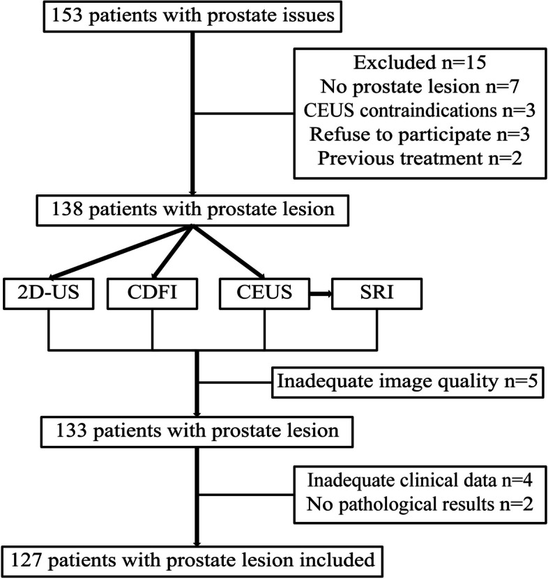
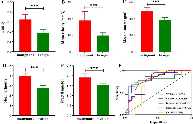
This study included 127 patients with prostate lesion who presented at Renmin Hospital of Wuhan University between November 2023 and June 2024 were included in this study. All the patients underwent transrectal US (TRUS), contrast-enhanced US (CEUS), and US SRI. CEUS parameters of time-intensity curve (TIC): arrival time (AT), rising time (RT), time to peak (TTP), peak intensity (PKI), falling time (FT), mean transit time (MTT), ascending slope (AS), descending slope (DS), D/A slope ratio (SR), and area under the TIC (AUC). US SRI parameters: microvessel density (MVD), microvessel diameter (D), microvessel velocity (V), microvessel tortuosity (T), and fractal number (FN), were analyzed and compared between prostate benign and malignant lesion.

RESULTS

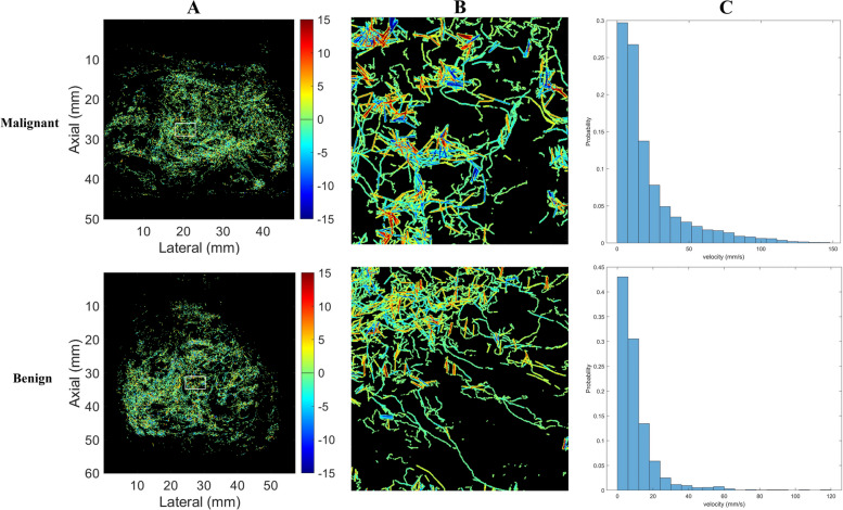
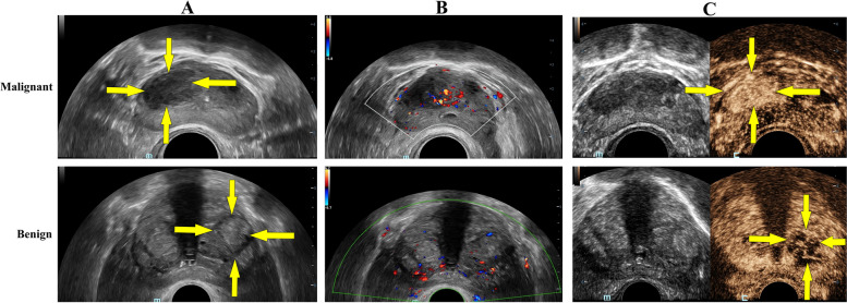
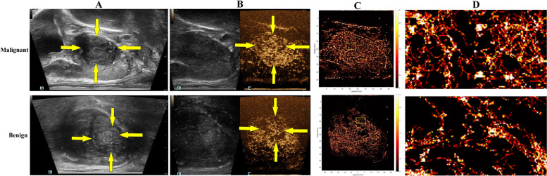
The tumor markers of prostate in the malignant group were all higher than those in the benign group, and the differences were statistically significant (P < 0.001). The TIC parameters of CEUS revealed that the PKI, AS, DS, and AUC were significantly higher in the malignant group than in the benign group (P < 0.001), whereas the RT, TTP and FT in the malignant group were significantly lower (P < 0.001). Malignant lesion exhibited significantly higher MVD, larger D, faster V, greater T, and more complex FN than benign lesion (P < 0.001).

CONCLUSIONS

US SRI is a promising non-invasive imaging modality that can provide detailed microvessel characteristics of prostate lesion, offering an advancement in the differential diagnosis for prostate lesion. And, US SRI may be a valuable tool in clinical practice with its ability to display and quantify microvessel with high precision.

摘要

背景

前列腺癌(PCa)是全球男性癌症相关发病和死亡的主要原因。早期准确诊断对于有效治疗和预后至关重要。传统的侵入性检查,如图像引导下的前列腺活检,常常会引起不适和并发症,导致一些患者不愿接受这些必要的检查。本研究旨在探讨使用超声超分辨率成像(US SRI)非侵入性评估前列腺病变微血管特征的可行性和临床价值。

方法

本研究纳入了2023年11月至2024年6月期间在武汉大学人民医院就诊的127例前列腺病变患者。所有患者均接受经直肠超声(TRUS)、超声造影(CEUS)和US SRI检查。分析并比较前列腺良性和恶性病变的CEUS时间强度曲线(TIC)参数:达峰时间(AT)、上升时间(RT)、峰值时间(TTP)、峰值强度(PKI)、下降时间(FT)、平均通过时间(MTT)、上升斜率(AS)、下降斜率(DS)、D/A斜率比(SR)以及TIC下面积(AUC)。US SRI参数:微血管密度(MVD)、微血管直径(D)、微血管速度(V)、微血管迂曲度(T)和分形维数(FN)。

结果

恶性组前列腺肿瘤标志物均高于良性组,差异具有统计学意义(P < 0.001)。CEUS的TIC参数显示,恶性组的PKI、AS、DS和AUC显著高于良性组(P < 0.001),而恶性组的RT、TTP和FT显著低于良性组(P < 0.001)。恶性病变的MVD、D、V、T和FN均显著高于良性病变(P < 0.001)。

结论

US SRI是一种有前景的非侵入性成像方式,能够提供前列腺病变详细的微血管特征,为前列腺病变的鉴别诊断带来进展。并且,US SRI能够高精度地显示和量化微血管,可能成为临床实践中的一种有价值的工具。

https://cdn.ncbi.nlm.nih.gov/pmc/blobs/6ee6/11706184/d7aebb260471/40644_2024_819_Fig5_HTML.jpg
https://cdn.ncbi.nlm.nih.gov/pmc/blobs/6ee6/11706184/89d42d7f11cd/40644_2024_819_Fig1_HTML.jpg
https://cdn.ncbi.nlm.nih.gov/pmc/blobs/6ee6/11706184/289394b16b6c/40644_2024_819_Fig2_HTML.jpg
https://cdn.ncbi.nlm.nih.gov/pmc/blobs/6ee6/11706184/8517a72039b3/40644_2024_819_Fig3_HTML.jpg
https://cdn.ncbi.nlm.nih.gov/pmc/blobs/6ee6/11706184/5fe184014b48/40644_2024_819_Fig4_HTML.jpg
https://cdn.ncbi.nlm.nih.gov/pmc/blobs/6ee6/11706184/d7aebb260471/40644_2024_819_Fig5_HTML.jpg

相似文献

1
Ultrasound super-resolution imaging for non-invasive assessment of microvessel in prostate lesion.超声超分辨率成像用于前列腺病变微血管的无创评估。
Cancer Imaging. 2025 Jan 7;25(1):1. doi: 10.1186/s40644-024-00819-z.
2
A Prospective Study on the Value of Ultrasound Microflow Assessment to Distinguish Malignant from Benign Solid Breast Masses: Association between Ultrasound Parameters and Histologic Microvessel Densities.超声微血管流评估对鉴别良恶性实性乳腺肿块的价值的前瞻性研究:超声参数与组织微血管密度的相关性。
Korean J Radiol. 2019 May;20(5):759-772. doi: 10.3348/kjr.2018.0515.
3
Correlation of lymph node metastasis with contrast-enhanced ultrasound features, microvessel density and microvessel area in patients with papillary thyroid carcinoma.甲状腺乳头状癌患者淋巴结转移与超声造影特征、微血管密度及微血管面积的相关性
Clin Hemorheol Microcirc. 2022;82(4):361-370. doi: 10.3233/CH-221545.
4
Contrast-enhanced ultrasonography for the detection and characterization of prostate cancer: correlation with microvessel density and Gleason score.超声造影在前列腺癌检测及分级中的应用:与微血管密度和 Gleason 评分的相关性。
Clin Radiol. 2011 Aug;66(8):732-7. doi: 10.1016/j.crad.2011.02.013. Epub 2011 Apr 23.
5
Multiparametric MRI in detection and staging of prostate cancer.多参数磁共振成像在前列腺癌检测与分期中的应用
Dan Med J. 2017 Feb;64(2).
6
Contrast-enhanced ultrasound evaluation of the prostate before transrectal ultrasound-guided biopsy can improve diagnostic sensitivity: A STARD-compliant article.经直肠超声引导下前列腺穿刺活检前的超声造影评估可提高诊断敏感性:一篇符合STARD标准的文章。
Medicine (Baltimore). 2020 May;99(19):e19946. doi: 10.1097/MD.0000000000019946.
7
Contrast enhanced ultrasound (CEUS) with MRI image fusion for monitoring focal therapy of prostate cancer with high intensity focused ultrasound (HIFU)1.对比增强超声(CEUS)与MRI图像融合用于监测高强度聚焦超声(HIFU)治疗前列腺癌的局部治疗1。
Clin Hemorheol Microcirc. 2018;69(1-2):93-100. doi: 10.3233/CH-189123.
8
Contrast-Enhanced Ultrasound Differentiation Between Low- and High- Grade Bladder Urothelial Carcinoma and Correlation With Tumor Microvessel Density.对比增强超声鉴别低级别和高级别膀胱尿路上皮癌及其与肿瘤微血管密度的相关性
J Ultrasound Med. 2017 Nov;36(11):2287-2297. doi: 10.1002/jum.14262. Epub 2017 May 27.
9
Contrast enhanced ultrasound (CEUS) with parametric imaging and time intensity curve analysis (TIC) for evaluation of the success of prostate arterial embolization (PAE) in cases of prostate hyperplasia.对比增强超声(CEUS)联合参数成像和时间强度曲线分析(TIC)用于评估前列腺增生症患者前列腺动脉栓塞术(PAE)的疗效。
Clin Hemorheol Microcirc. 2020;76(2):143-153. doi: 10.3233/CH-209202.
10
Machine learning based on radiomics features combing B-mode transrectal ultrasound and contrast-enhanced ultrasound to improve peripheral zone prostate cancer detection.基于放射组学特征的机器学习结合 B 型经直肠超声和对比增强超声提高外周带前列腺癌检测能力。
Abdom Radiol (NY). 2024 Jan;49(1):141-150. doi: 10.1007/s00261-023-04050-5. Epub 2023 Oct 5.

本文引用的文献

1
[Cancer incidence and mortality in China, 2022].[2022年中国癌症发病率和死亡率]
Zhonghua Zhong Liu Za Zhi. 2024 Mar 23;46(3):221-231. doi: 10.3760/cma.j.cn112152-20240119-00035.
2
Cancer statistics, 2024.2024年癌症统计数据。
CA Cancer J Clin. 2024 Jan-Feb;74(1):12-49. doi: 10.3322/caac.21820. Epub 2024 Jan 17.
3
Acceleration-Based Kalman Tracking for Super-Resolution Ultrasound Imaging In Vivo.基于加速度的超分辨率超声体内成像的卡尔曼跟踪。
IEEE Trans Ultrason Ferroelectr Freq Control. 2023 Dec;70(12):1739-1748. doi: 10.1109/TUFFC.2023.3326863. Epub 2023 Dec 14.
4
Quantitative tissue perfusion imaging using nonlinear ultrasound localization microscopy.利用非线性超声定位显微镜进行定量组织灌注成像。
Sci Rep. 2022 Dec 19;12(1):21943. doi: 10.1038/s41598-022-24986-w.
5
Ultrasound super-resolution imaging for differential diagnosis of breast masses.用于乳腺肿块鉴别诊断的超声超分辨率成像
Front Oncol. 2022 Nov 3;12:1049991. doi: 10.3389/fonc.2022.1049991. eCollection 2022.
6
Ultrasound super-resolution imaging for the differential diagnosis of thyroid nodules: A pilot study.超声超分辨率成像在甲状腺结节鉴别诊断中的初步研究
Front Oncol. 2022 Oct 27;12:978164. doi: 10.3389/fonc.2022.978164. eCollection 2022.
7
Clinically Localized Prostate Cancer: AUA/ASTRO Guideline.临床局限性前列腺癌:美国泌尿外科学会/美国放射肿瘤学会指南
J Urol. 2022 Sep;208(3):505-507. doi: 10.1097/JU.0000000000002854. Epub 2022 Jul 5.
8
A Review of Clinical Applications for Super-resolution Ultrasound Localization Microscopy.超分辨率超声定位显微镜的临床应用综述
Curr Med Sci. 2022 Feb;42(1):1-16. doi: 10.1007/s11596-021-2459-2. Epub 2022 Feb 15.
9
Baseline microvessel density predicts response to neoadjuvant bevacizumab treatment of locally advanced breast cancer.基线微血管密度可预测局部晚期乳腺癌新辅助贝伐单抗治疗的反应。
Sci Rep. 2021 Feb 9;11(1):3388. doi: 10.1038/s41598-021-81914-0.
10
EAU-EANM-ESTRO-ESUR-SIOG Guidelines on Prostate Cancer-2020 Update. Part 1: Screening, Diagnosis, and Local Treatment with Curative Intent.EAU-EANM-ESTRO-ESUR-SIOG 前列腺癌指南-2020 版更新。第 1 部分:筛查、诊断和以治愈为目的的局部治疗。
Eur Urol. 2021 Feb;79(2):243-262. doi: 10.1016/j.eururo.2020.09.042. Epub 2020 Nov 7.