Rosli Salwa, Abd Halim Haizlene, Md-Yasin Mazapuspavina, Abu Bakar Nur Aini
Department of Primary Care Medicine, Faculty of Universiti Teknologi MARA (UiTM), Sungai Buloh, Selangor, Malaysia.
Hospital Al-Sultan Abdullah (HASA), Universiti Teknologi MARA (UiTM), Puncak Alam, Selangor, Malaysia.
Am J Case Rep. 2025 Jan 7;26:e945897. doi: 10.12659/AJCR.945897.
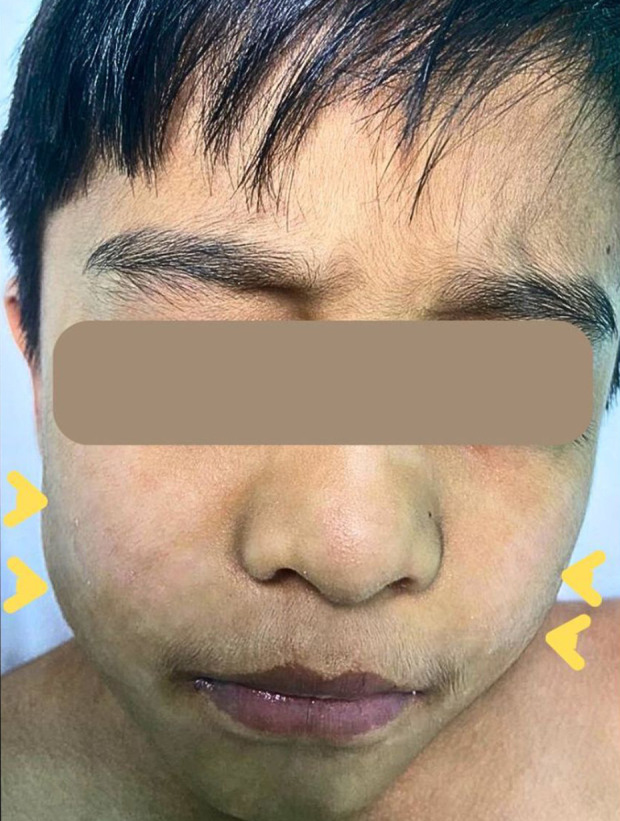
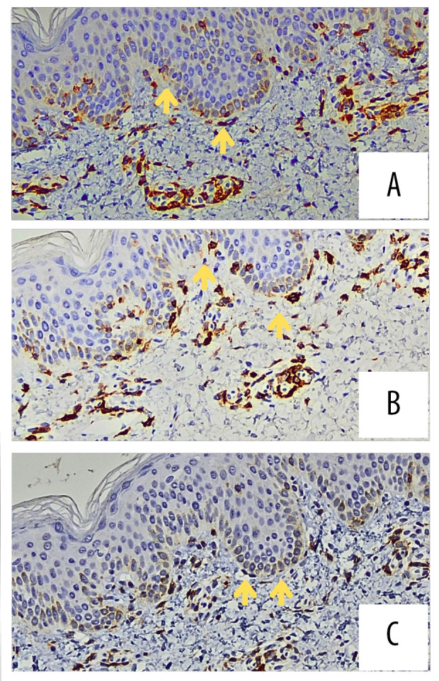
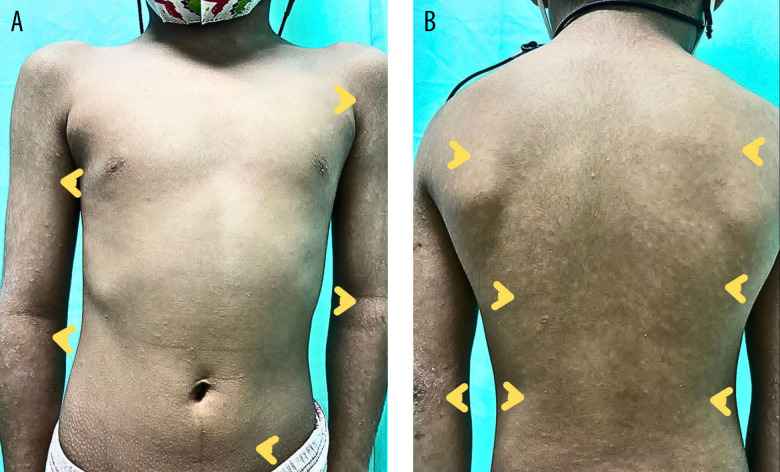
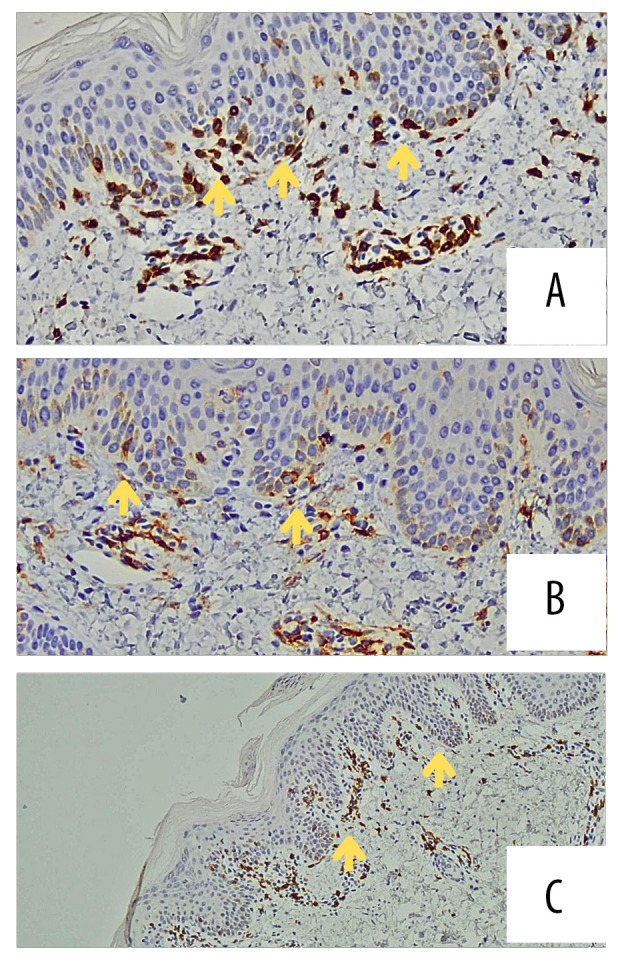
BACKGROUND Primary cutaneous lymphomas (PCL) are a multifaceted spectrum of cutaneous T cell lymphoma (CTCL) and cutaneous B cell lymphomas (CBCL). Mycosis fungoides (MF) is a rare subset of CTCL that primarily affects adults, and its occurrence in children is exceedingly rare. Most pediatric MF manifests as hypopigmented patches resembling other benign dermatoses, causing diagnostic challenges. This report outlines a case of pediatric MF in a 7-year-old Malaysian boy. CASE REPORT A 7-year-old boy exhibited progressing skin lesions characterized initially by erythematous, papular rashes over the face and upper limbs, then to the whole body, becoming hypopigmented, with pruritus and scaling for 1 year. Multiple clinics treated him for eczema and pityriasis alba but he responded poorly to courses of various topical steroids and emollient treatment. Due to the refractory nature of the lesions, he was subsequently referred to a dermatology clinic, where 2 skin biopsies were performed. The first biopsy revealed epidermotropism of atypical lymphocytes, consistent with MF. Immunohistochemical analysis revealed positive CD3+ expression with slightly reduced CD4+, CD7+, and CD8+ expression, and normal CD2+ and CD5+ expression at the epidermis level. Nevertheless, due to the rarity of MF in children, a second biopsy was performed, validating the diagnosis. CONCLUSIONS Pediatric MF is a rare and challenging diagnosis. This case report highlights the importance of close monitoring of unresolved hypopigmented lesions and increased vigilance on lesions not responding to standard treatment. Timely diagnosis with support of skin biopsy is crucial to avoid potentially serious disease progression and helps provide appropriate management leading to improved outcomes.
背景 原发性皮肤淋巴瘤(PCL)是皮肤T细胞淋巴瘤(CTCL)和皮肤B细胞淋巴瘤(CBCL)的一个多方面的谱系。蕈样肉芽肿(MF)是CTCL的一个罕见亚型,主要影响成年人,在儿童中极为罕见。大多数儿童MF表现为色素减退斑,类似于其他良性皮肤病,这给诊断带来了挑战。本报告概述了一名7岁马来西亚男孩患儿童MF的病例。病例报告 一名7岁男孩出现进行性皮肤病变,最初表现为面部和上肢的红斑、丘疹性皮疹,随后发展至全身,变为色素减退,伴有瘙痒和鳞屑,持续1年。多家诊所对他进行了湿疹和白色糠疹的治疗,但他对各种外用类固醇和润肤剂治疗方案反应不佳。由于病变的难治性,他随后被转诊至皮肤科诊所,在那里进行了2次皮肤活检。第一次活检显示非典型淋巴细胞向表皮浸润,符合MF。免疫组织化学分析显示在表皮水平CD3 + 表达阳性,CD4 +、CD7 + 和CD8 + 表达略有降低,CD2 + 和CD5 + 表达正常。然而,由于儿童MF罕见,进行了第二次活检以确诊。结论 儿童MF是一种罕见且具有挑战性的诊断。本病例报告强调了密切监测未消退的色素减退性病变以及对标准治疗无反应的病变提高警惕的重要性。在皮肤活检的支持下及时诊断对于避免潜在的严重疾病进展至关重要,并有助于提供适当的管理以改善预后。