Nardi G, Torcq L, Schmidt A A, Olivo-Marin J-C
Biological Image Analysis Unit, Institut Pasteur, Université Paris Cité, Paris, France.
CNRS UMR3691, Paris, France.
Biol Imaging. 2024 Nov 11;4:e11. doi: 10.1017/S2633903X24000102. eCollection 2024.
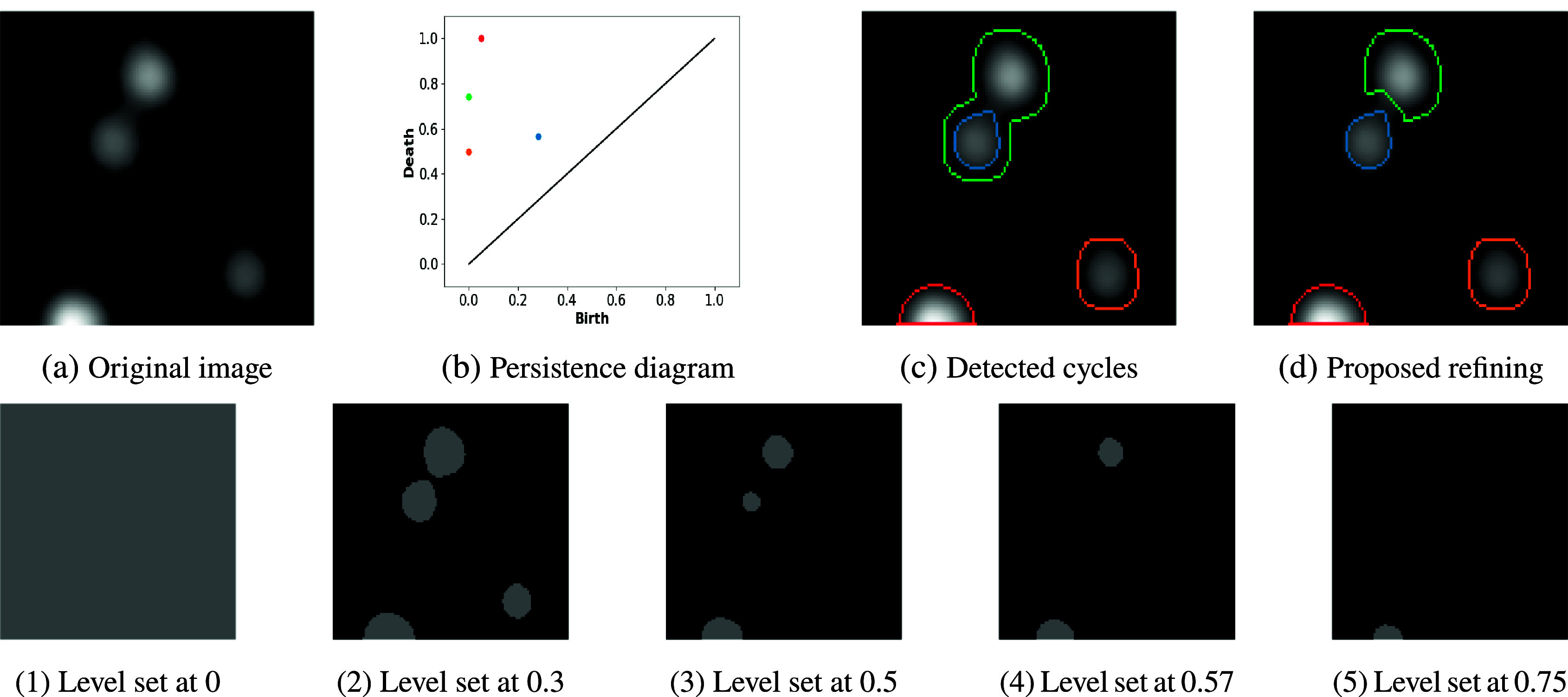
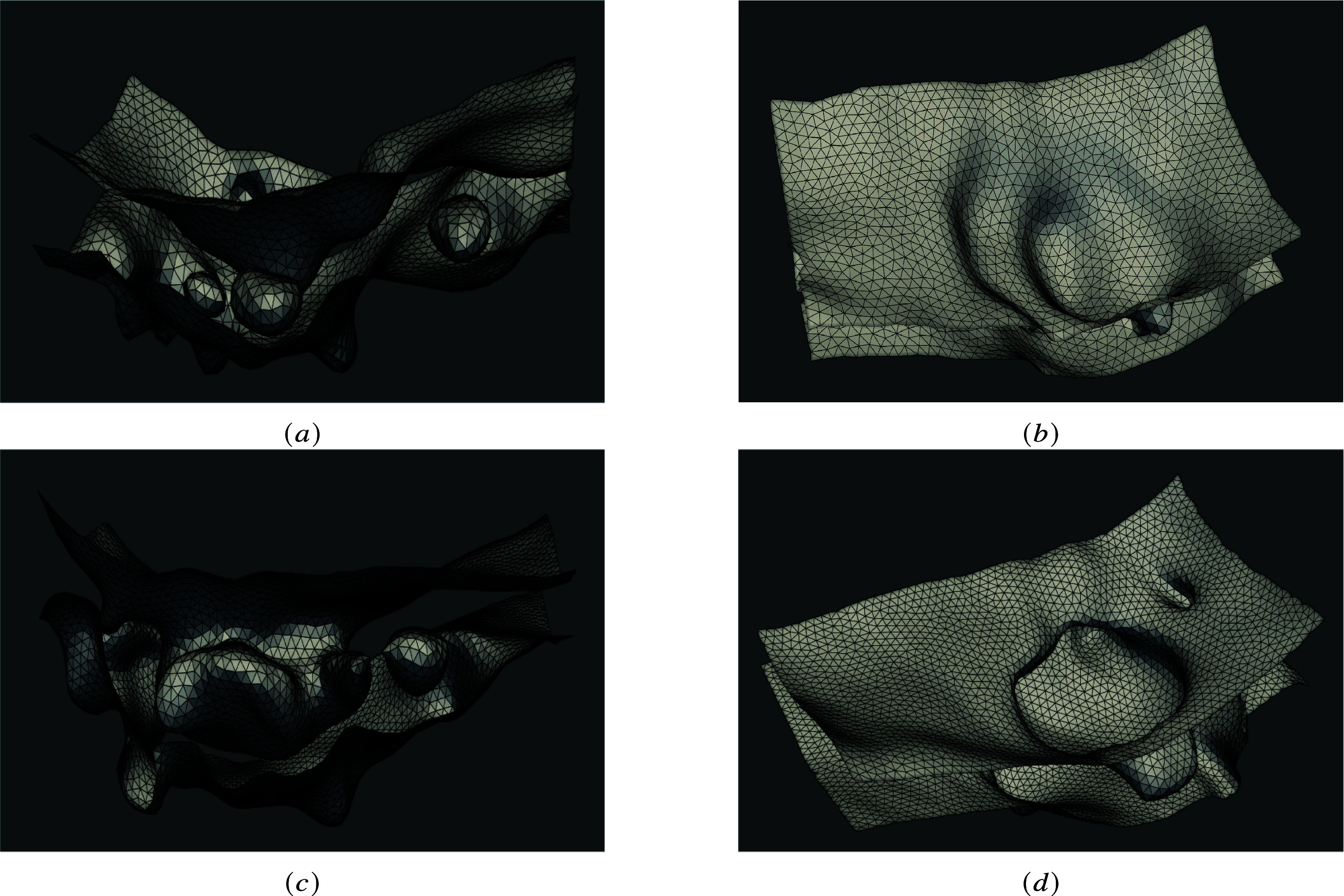
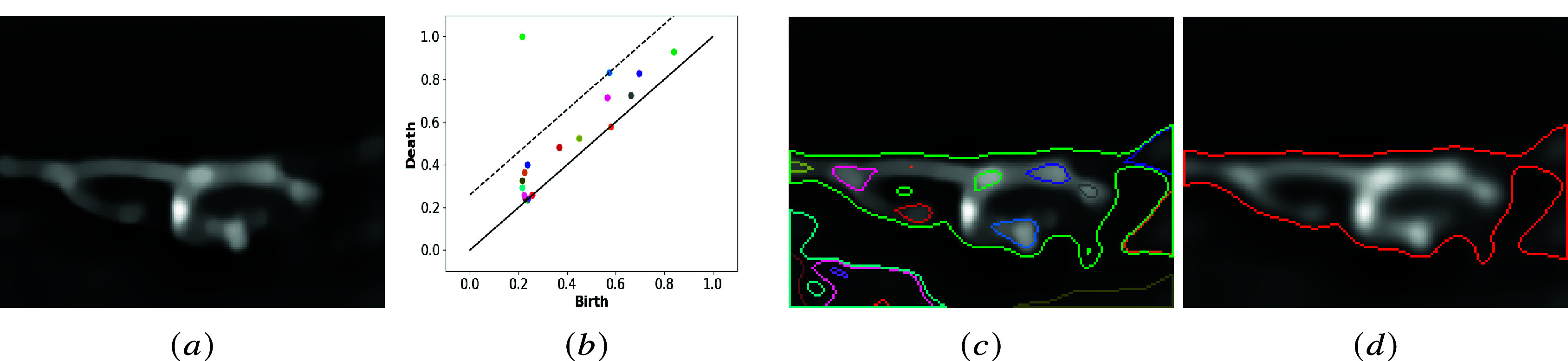
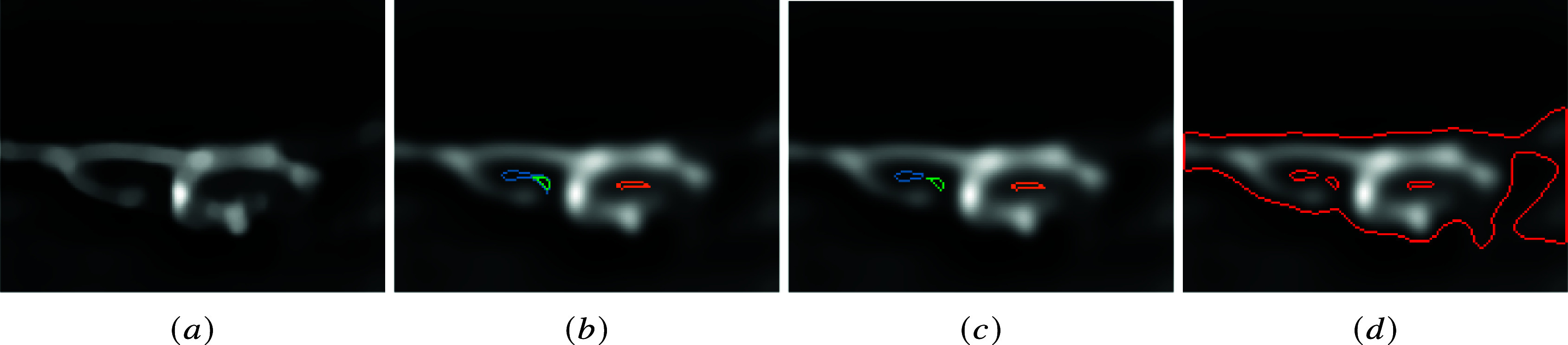
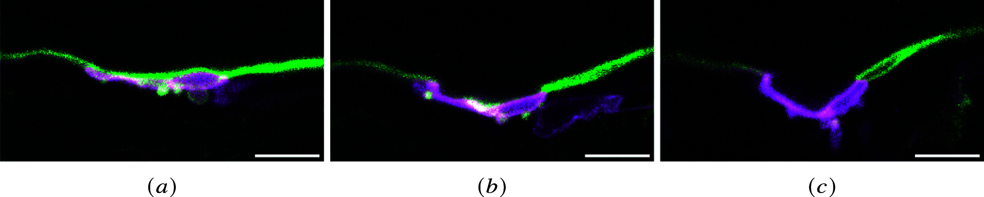
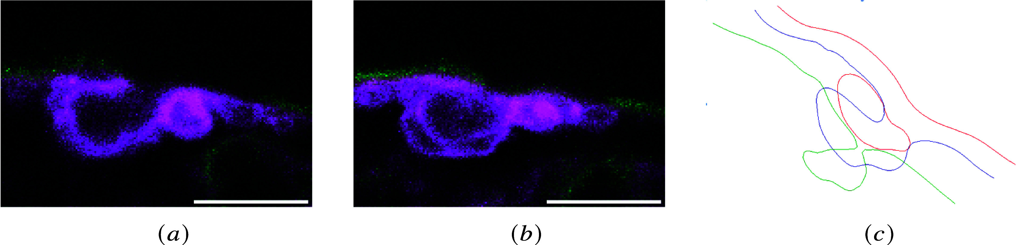
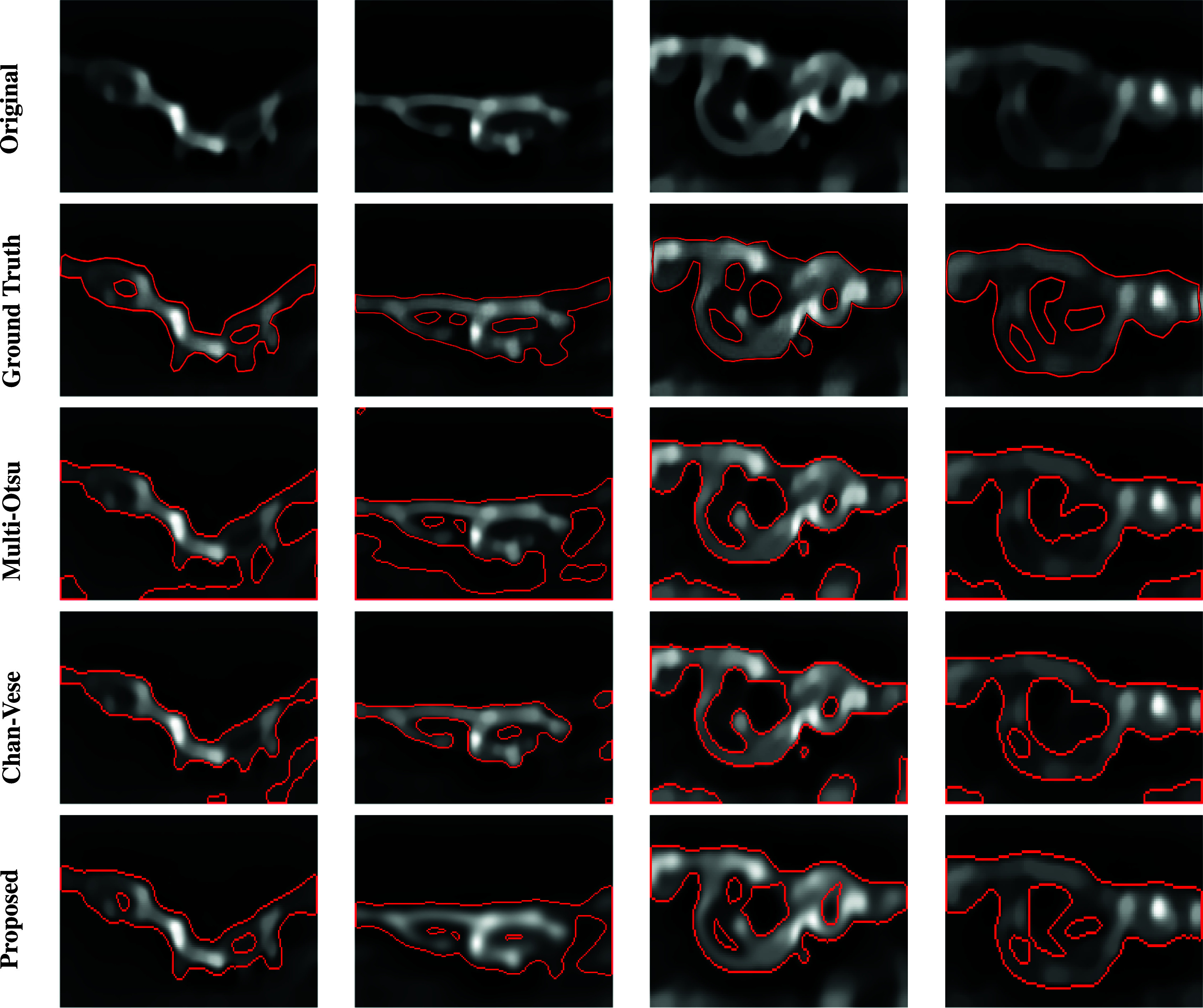
We develop a novel method for image segmentation of 3D confocal microscopy images of emerging hematopoietic stem cells. The method is based on the theory of persistent homology and uses an optimal threshold to select the most persistent cycles in the persistence diagram. This enables the segmentation of the image's most contrasted and representative shapes. Coupling this segmentation method with a meshing algorithm, we define a pipeline for 3D reconstruction of confocal volumes. Compared to related methods, this approach improves shape segmentation, is more ergonomic to automatize, and has fewer parameters. We apply it to the segmentation of membranes, at subcellular resolution, of cells involved in the endothelial-to-hematopoietic transition (EHT) in the zebrafish embryos.
我们开发了一种用于新兴造血干细胞三维共聚焦显微镜图像分割的新方法。该方法基于持久同调理论,并使用最优阈值在持久图中选择最持久的圈。这使得能够分割出图像中对比度最高且最具代表性的形状。将这种分割方法与网格划分算法相结合,我们定义了一个用于共聚焦体积三维重建的流程。与相关方法相比,这种方法改进了形状分割,更便于自动化操作,且参数更少。我们将其应用于斑马鱼胚胎中参与内皮向造血转变(EHT)的细胞亚细胞分辨率下的膜分割。