• 文献检索
  • 文档翻译
  • 深度研究
  • 学术资讯
  • Suppr Zotero 插件Zotero 插件
  • 邀请有礼
  • 套餐&价格
  • 历史记录
应用&插件
Suppr Zotero 插件Zotero 插件浏览器插件Mac 客户端Windows 客户端微信小程序
定价
高级版会员购买积分包购买API积分包
服务
文献检索文档翻译深度研究API 文档MCP 服务
关于我们
关于 Suppr公司介绍联系我们用户协议隐私条款
关注我们

Suppr 超能文献

核心技术专利:CN118964589B侵权必究
粤ICP备2023148730 号-1Suppr @ 2026

文献检索

告别复杂PubMed语法,用中文像聊天一样搜索,搜遍4000万医学文献。AI智能推荐,让科研检索更轻松。

立即免费搜索

文件翻译

保留排版,准确专业,支持PDF/Word/PPT等文件格式,支持 12+语言互译。

免费翻译文档

深度研究

AI帮你快速写综述,25分钟生成高质量综述,智能提取关键信息,辅助科研写作。

立即免费体验

用于目标检测的拓扑图像修改和皮肤损伤的拓扑图像处理。

Topological image modification for object detection and topological image processing of skin lesions.

机构信息

Stanford Center for Biomedical Informatics Research (BMIR), Department of Medicine, Stanford University School of Medicine, Stanford, CA, 94305, USA.

IDLab, Department of Electronics and Information Systems, Ghent University, Technologiepark-Zwijnaarde 19, 9052, Gent, Belgium.

出版信息

Sci Rep. 2020 Dec 3;10(1):21061. doi: 10.1038/s41598-020-77933-y.

DOI:10.1038/s41598-020-77933-y
PMID:33273628
原文链接:https://pmc.ncbi.nlm.nih.gov/articles/PMC7713118/
Abstract

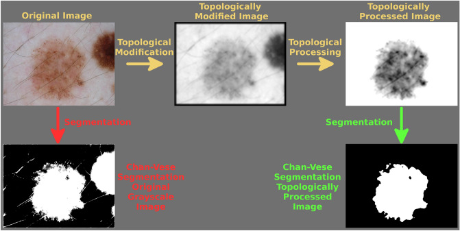
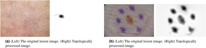
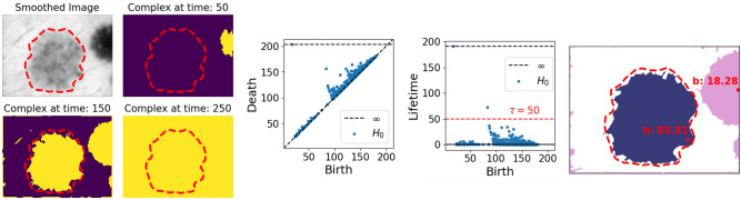
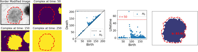
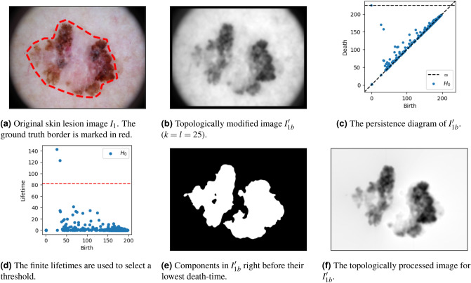
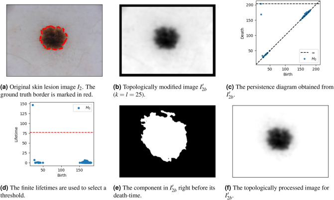
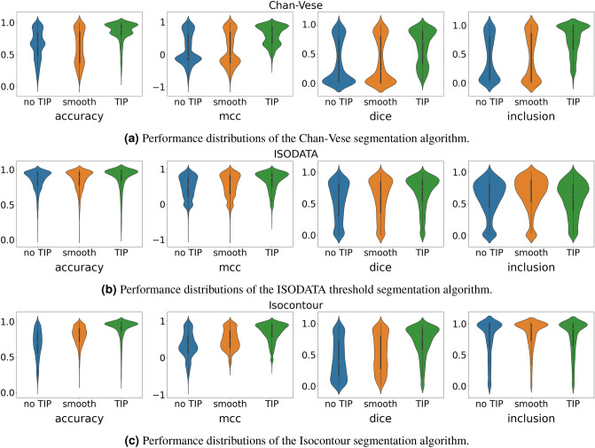
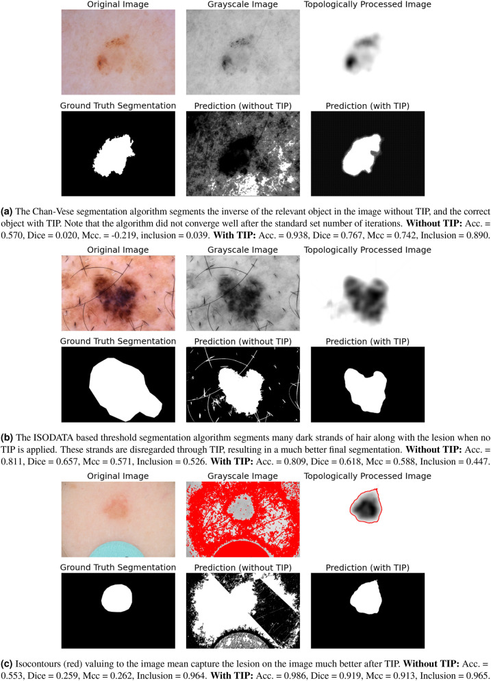
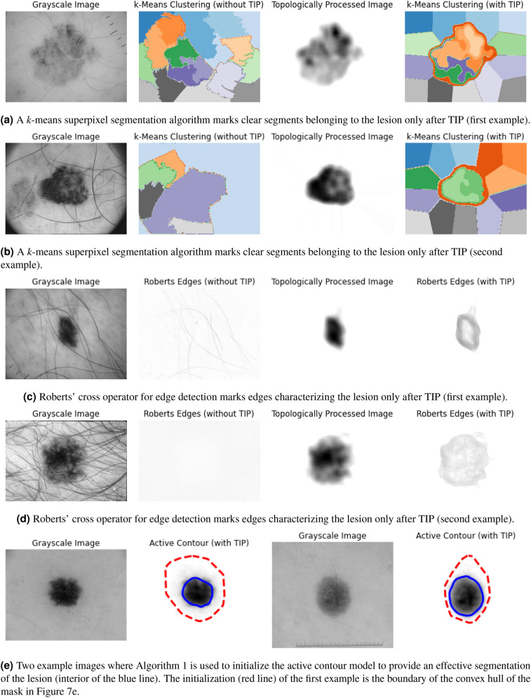
We propose a new method based on Topological Data Analysis (TDA) consisting of Topological Image Modification (TIM) and Topological Image Processing (TIP) for object detection. Through this newly introduced method, we artificially destruct irrelevant objects, and construct new objects with known topological properties in irrelevant regions of an image. This ensures that we are able to identify the important objects in relevant regions of the image. We do this by means of persistent homology, which allows us to simultaneously select appropriate thresholds, as well as the objects corresponding to these thresholds, and separate them from the noisy background of an image. This leads to a new image, processed in a completely unsupervised manner, from which one may more efficiently extract important objects. We demonstrate the usefulness of this proposed method for topological image processing through a case-study of unsupervised segmentation of the ISIC 2018 skin lesion images. Code for this project is available on https://bitbucket.org/ghentdatascience/topimgprocess .

摘要

我们提出了一种基于拓扑数据分析(TDA)的新方法,包括拓扑图像修改(TIM)和拓扑图像处理(TIP),用于目标检测。通过这种新引入的方法,我们人为地破坏不相关的物体,并在图像的不相关区域中用具有已知拓扑性质的新物体来构造。这确保了我们能够识别图像相关区域中的重要物体。我们通过持久同调来做到这一点,这使我们能够同时选择合适的阈值,以及与这些阈值对应的物体,并将它们与图像的噪声背景区分开来。这导致了一个新的图像,以完全无监督的方式进行处理,从中可以更有效地提取重要的物体。我们通过对 ISIC 2018 皮肤病变图像的无监督分割的案例研究,展示了这种拓扑图像处理方法的有用性。该项目的代码可在 https://bitbucket.org/ghentdatascience/topimgprocess 上获得。

https://cdn.ncbi.nlm.nih.gov/pmc/blobs/a2c3/7713118/ee76cac349da/41598_2020_77933_Fig11_HTML.jpg
https://cdn.ncbi.nlm.nih.gov/pmc/blobs/a2c3/7713118/403b36d52f5e/41598_2020_77933_Fig1_HTML.jpg
https://cdn.ncbi.nlm.nih.gov/pmc/blobs/a2c3/7713118/ee68dbc72d90/41598_2020_77933_Fig2_HTML.jpg
https://cdn.ncbi.nlm.nih.gov/pmc/blobs/a2c3/7713118/c221bc6c59e8/41598_2020_77933_Fig3_HTML.jpg
https://cdn.ncbi.nlm.nih.gov/pmc/blobs/a2c3/7713118/685059190ca3/41598_2020_77933_Fig4_HTML.jpg
https://cdn.ncbi.nlm.nih.gov/pmc/blobs/a2c3/7713118/98bd20b61332/41598_2020_77933_Fig5_HTML.jpg
https://cdn.ncbi.nlm.nih.gov/pmc/blobs/a2c3/7713118/e272599555c7/41598_2020_77933_Fig6_HTML.jpg
https://cdn.ncbi.nlm.nih.gov/pmc/blobs/a2c3/7713118/5cc6ac4ade8a/41598_2020_77933_Fig7_HTML.jpg
https://cdn.ncbi.nlm.nih.gov/pmc/blobs/a2c3/7713118/16ed1517da10/41598_2020_77933_Fig8_HTML.jpg
https://cdn.ncbi.nlm.nih.gov/pmc/blobs/a2c3/7713118/a6b758528742/41598_2020_77933_Fig9_HTML.jpg
https://cdn.ncbi.nlm.nih.gov/pmc/blobs/a2c3/7713118/fca15ad6ae27/41598_2020_77933_Fig10_HTML.jpg
https://cdn.ncbi.nlm.nih.gov/pmc/blobs/a2c3/7713118/ee76cac349da/41598_2020_77933_Fig11_HTML.jpg

相似文献

1
Topological image modification for object detection and topological image processing of skin lesions.用于目标检测的拓扑图像修改和皮肤损伤的拓扑图像处理。
Sci Rep. 2020 Dec 3;10(1):21061. doi: 10.1038/s41598-020-77933-y.
2
Efficient skin lesion segmentation using separable-Unet with stochastic weight averaging.使用可分离 U-Net 和随机权重平均化实现高效的皮肤病变分割。
Comput Methods Programs Biomed. 2019 Sep;178:289-301. doi: 10.1016/j.cmpb.2019.07.005. Epub 2019 Jul 8.
3
Robust detection and segmentation of cell nuclei in biomedical images based on a computational topology framework.基于计算拓扑学框架的生物医学图像中细胞核的稳健检测和分割。
Med Image Anal. 2017 May;38:90-103. doi: 10.1016/j.media.2017.02.009. Epub 2017 Mar 6.
4
Fast processing of microscopic images using object-based extended depth of field.使用基于对象的扩展景深快速处理显微图像。
BMC Bioinformatics. 2016 Dec 22;17(Suppl 19):516. doi: 10.1186/s12859-016-1373-2.
5
Segmentation of biomedical images based on a computational topology framework.基于计算拓扑学框架的生物医学图像分割。
Semin Immunol. 2020 Apr;48:101432. doi: 10.1016/j.smim.2020.101432. Epub 2020 Dec 2.
6
Topological equivalence between a 3D object and the reconstruction of its digital image.三维物体与其数字图像重建之间的拓扑等价性。
IEEE Trans Pattern Anal Mach Intell. 2007 Jan;29(1):126-40. doi: 10.1109/tpami.2007.250604.
7
Integrating watersheds and critical point analysis for object detection in discrete 2D images.在离散二维图像中集成流域分割和临界点分析以进行目标检测。
Med Image Anal. 2004 Sep;8(3):177-85. doi: 10.1016/j.media.2004.06.002.
8
Skin Lesion Segmentation from Dermoscopic Images Using Convolutional Neural Network.基于卷积神经网络的皮肤镜图像皮损分割。
Sensors (Basel). 2020 Mar 13;20(6):1601. doi: 10.3390/s20061601.
9
Structure and location preserving topological representation with applications on CT segmentation.具有结构和位置保持性的拓扑表示及其在CT分割中的应用
Annu Int Conf IEEE Eng Med Biol Soc. 2017 Jul;2017:548-551. doi: 10.1109/EMBC.2017.8036883.
10
Unsupervised category modeling, recognition, and segmentation in images.图像中的无监督类别建模、识别与分割。
IEEE Trans Pattern Anal Mach Intell. 2008 Dec;30(12):2158-74. doi: 10.1109/TPAMI.2008.24.

引用本文的文献

1
AFTG-Net: A Deep Attention-based Fusion Framework of Topological and Gradient Features for Pathological Image Analysis.AFTG-Net:一种基于深度注意力的拓扑和梯度特征融合框架,用于病理图像分析。
Res Sq. 2025 Jul 11:rs.3.rs-6710077. doi: 10.21203/rs.3.rs-6710077/v1.
2
Topology-based segmentation of 3D confocal images of emerging hematopoietic stem cells in the zebrafish embryo.基于拓扑学的斑马鱼胚胎中造血干细胞出现的三维共聚焦图像分割
Biol Imaging. 2024 Nov 11;4:e11. doi: 10.1017/S2633903X24000102. eCollection 2024.
3
Scoping Review: Methods and Applications of Spatial Transcriptomics in Tumor Research.

本文引用的文献

1
Marking the Path Toward Artificial Intelligence-Based Image Classification in Dermatology.迈向皮肤科基于人工智能的图像分类之路
JAMA Dermatol. 2019 Oct 1;155(10):1105-1106. doi: 10.1001/jamadermatol.2019.1633.
2
Topological data analysis of high resolution diabetic retinopathy images.高分辨率糖尿病视网膜病变图像的拓扑数据分析。
PLoS One. 2019 May 24;14(5):e0217413. doi: 10.1371/journal.pone.0217413. eCollection 2019.
3
Artificial intelligence and dermatology: opportunities, challenges, and future directions.人工智能与皮肤病学:机遇、挑战及未来方向。
综述:空间转录组学在肿瘤研究中的方法与应用
Cancers (Basel). 2024 Sep 6;16(17):3100. doi: 10.3390/cancers16173100.
4
Advancing Precision Medicine: Algebraic Topology and Differential Geometry in Radiology and Computational Pathology.推进精准医学:放射学和计算病理学中的代数拓扑和微分几何。
Lab Invest. 2024 Jun;104(6):102060. doi: 10.1016/j.labinv.2024.102060. Epub 2024 Apr 16.
5
PrestoCell: A persistence-based clustering approach for rapid and robust segmentation of cellular morphology in three-dimensional data.PrestoCell:一种基于持久性的聚类方法,用于快速、稳健地分割三维数据中的细胞形态。
PLoS One. 2024 Feb 29;19(2):e0299006. doi: 10.1371/journal.pone.0299006. eCollection 2024.
6
Artificial Intelligence Algorithms for Benign vs. Malignant Dermoscopic Skin Lesion Image Classification.用于良性与恶性皮肤镜下皮肤病变图像分类的人工智能算法
Bioengineering (Basel). 2023 Nov 16;10(11):1322. doi: 10.3390/bioengineering10111322.
7
Topological data analysis of thoracic radiographic images shows improved radiomics-based lung tumor histology prediction.胸部X光影像的拓扑数据分析显示基于影像组学的肺肿瘤组织学预测得到改善。
Patterns (N Y). 2022 Dec 12;4(1):100657. doi: 10.1016/j.patter.2022.100657. eCollection 2023 Jan 13.
Semin Cutan Med Surg. 2019 Mar 1;38(1):E31-37. doi: 10.12788/j.sder.2019..
4
The HAM10000 dataset, a large collection of multi-source dermatoscopic images of common pigmented skin lesions.HAM10000 数据集,一个大型的常见色素性皮肤病变多源皮肤镜图像集合。
Sci Data. 2018 Aug 14;5:180161. doi: 10.1038/sdata.2018.161.
5
SLIC superpixels compared to state-of-the-art superpixel methods.SLIC 超像素与最先进的超像素方法比较。
IEEE Trans Pattern Anal Mach Intell. 2012 Nov;34(11):2274-82. doi: 10.1109/TPAMI.2012.120.