• 文献检索
  • 文档翻译
  • 深度研究
  • 学术资讯
  • Suppr Zotero 插件Zotero 插件
  • 邀请有礼
  • 套餐&价格
  • 历史记录
应用&插件
Suppr Zotero 插件Zotero 插件浏览器插件Mac 客户端Windows 客户端微信小程序
定价
高级版会员购买积分包购买API积分包
服务
文献检索文档翻译深度研究API 文档MCP 服务
关于我们
关于 Suppr公司介绍联系我们用户协议隐私条款
关注我们

Suppr 超能文献

核心技术专利:CN118964589B侵权必究
粤ICP备2023148730 号-1Suppr @ 2026

文献检索

告别复杂PubMed语法,用中文像聊天一样搜索,搜遍4000万医学文献。AI智能推荐,让科研检索更轻松。

立即免费搜索

文件翻译

保留排版,准确专业,支持PDF/Word/PPT等文件格式,支持 12+语言互译。

免费翻译文档

深度研究

AI帮你快速写综述,25分钟生成高质量综述,智能提取关键信息,辅助科研写作。

立即免费体验

神经囊尾蚴病:解开影像学谜团

Neurocysticercosis: unwinding the radiological conundrum.

作者信息

Goddu Govindappa Sharath Kumar, Patil Megharanjini, Adiga Chaitra Parameshwara, Kumar Sujit, Goolahally Lakshmikanth N, Kumar Savith, Mathew Thomas

机构信息

Apollo Hospitals, Sheshadripuram, Bangalore, India.

Karnataka Institute of Medical Sciences, Hubli, India.

出版信息

Pol J Radiol. 2024 Nov 28;89:e549-e560. doi: 10.5114/pjr/193968. eCollection 2024.

DOI:10.5114/pjr/193968
PMID:39777324
原文链接:https://pmc.ncbi.nlm.nih.gov/articles/PMC11705042/
Abstract

PURPOSE

To study the distinct imaging characteristics of parenchymal neurocysticercosis (NCC) that aid in distinguishing it from other diseases.

MATERIAL AND METHODS

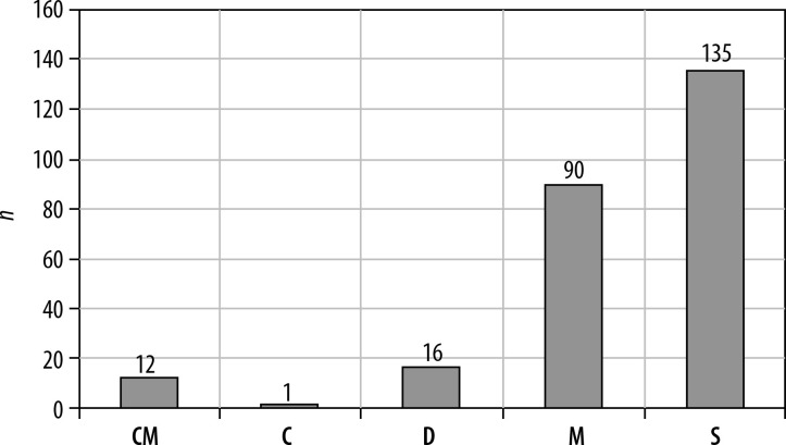
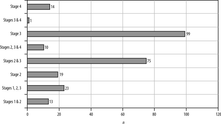
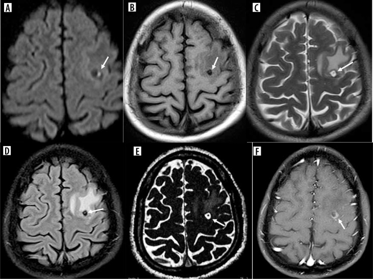
Two hundred fifty patients with NCC were selected based on identification of the scolex. T2 weighted, T1 fluid attenuated inversion recovery (FLAIR), T2 FLAIR, susceptibility weighted imaging, constructive interference in steady state, diffusion weighted imaging, and T1 weighted contrast sequences were performed. The imaging characteristics of the cysts at various stages were analyzed.

RESULTS

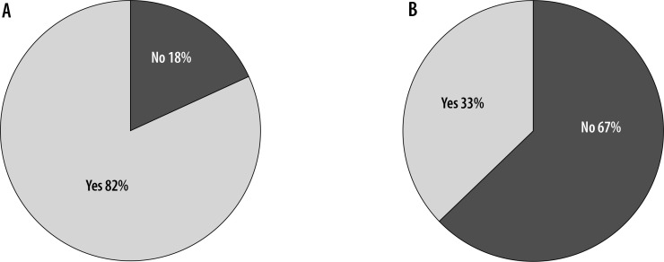
This study presents previously undocumented imaging findings of NCC, establishing absolute, major, and minor criteria for its diagnosis based on magnetic resonance imaging features.

CONCLUSIONS

Utilizing imaging criteria for diagnosing NCC, as per the study's sequences, can effectively narrow down differential diagnoses and lead to accurate identification.

摘要

目的

研究实质性神经囊尾蚴病(NCC)的独特影像学特征,以助于将其与其他疾病区分开来。

材料与方法

基于头节的识别选择了250例NCC患者。进行了T2加权、T1液体衰减反转恢复(FLAIR)、T2 FLAIR、磁敏感加权成像、稳态构成性干扰、扩散加权成像和T1加权对比序列检查。分析了囊肿在各个阶段的影像学特征。

结果

本研究呈现了NCC此前未记录的影像学表现,基于磁共振成像特征建立了其诊断的绝对、主要和次要标准。

结论

根据本研究的序列,利用影像学标准诊断NCC可有效缩小鉴别诊断范围并实现准确识别。

https://cdn.ncbi.nlm.nih.gov/pmc/blobs/43ed/11705042/6d000d92ecca/PJR-89-288904-g015.jpg
https://cdn.ncbi.nlm.nih.gov/pmc/blobs/43ed/11705042/65ee4865b9d9/PJR-89-288904-g001.jpg
https://cdn.ncbi.nlm.nih.gov/pmc/blobs/43ed/11705042/77cbf8e03d1f/PJR-89-288904-g002.jpg
https://cdn.ncbi.nlm.nih.gov/pmc/blobs/43ed/11705042/26cba2fc2f90/PJR-89-288904-g003.jpg
https://cdn.ncbi.nlm.nih.gov/pmc/blobs/43ed/11705042/7c032a663e2b/PJR-89-288904-g004.jpg
https://cdn.ncbi.nlm.nih.gov/pmc/blobs/43ed/11705042/6aa4c78ecf32/PJR-89-288904-g005.jpg
https://cdn.ncbi.nlm.nih.gov/pmc/blobs/43ed/11705042/f880d69808e7/PJR-89-288904-g006.jpg
https://cdn.ncbi.nlm.nih.gov/pmc/blobs/43ed/11705042/d504761dbe4d/PJR-89-288904-g007.jpg
https://cdn.ncbi.nlm.nih.gov/pmc/blobs/43ed/11705042/830b21c52658/PJR-89-288904-g008.jpg
https://cdn.ncbi.nlm.nih.gov/pmc/blobs/43ed/11705042/286dcb529c84/PJR-89-288904-g009.jpg
https://cdn.ncbi.nlm.nih.gov/pmc/blobs/43ed/11705042/9b49c7840931/PJR-89-288904-g010.jpg
https://cdn.ncbi.nlm.nih.gov/pmc/blobs/43ed/11705042/6fe996f5676e/PJR-89-288904-g011.jpg
https://cdn.ncbi.nlm.nih.gov/pmc/blobs/43ed/11705042/a3831a408d06/PJR-89-288904-g012.jpg
https://cdn.ncbi.nlm.nih.gov/pmc/blobs/43ed/11705042/70d3e91e0d16/PJR-89-288904-g013.jpg
https://cdn.ncbi.nlm.nih.gov/pmc/blobs/43ed/11705042/a05561d1710f/PJR-89-288904-g014.jpg
https://cdn.ncbi.nlm.nih.gov/pmc/blobs/43ed/11705042/6d000d92ecca/PJR-89-288904-g015.jpg

相似文献

1
Neurocysticercosis: unwinding the radiological conundrum.神经囊尾蚴病:解开影像学谜团
Pol J Radiol. 2024 Nov 28;89:e549-e560. doi: 10.5114/pjr/193968. eCollection 2024.
2
Newer magnetic resonance imaging techniques in neurocysticercosis.神经囊虫病的新型磁共振成像技术。
Neuroradiol J. 2020 Dec;33(6):538-544. doi: 10.1177/1971400920949394. Epub 2020 Aug 26.
3
Relevance of 3D magnetic resonance imaging sequences in diagnosing basal subarachnoid neurocysticercosis.三维磁共振成像序列在诊断基底池蛛网膜下腔神经囊尾蚴病中的相关性
Acta Trop. 2015 Dec;152:60-65. doi: 10.1016/j.actatropica.2015.08.017. Epub 2015 Aug 29.
4
Role of Multiparametric Magnetic Resonance Imaging of the Brain in Differentiating Neurocysticercosis From Tuberculoma.脑部多参数磁共振成像在鉴别神经囊尾蚴病和结核瘤中的作用
Cureus. 2023 May 14;15(5):e39003. doi: 10.7759/cureus.39003. eCollection 2023 May.
5
The role of conventional MR imaging sequences in the evaluation of neurocysticercosis: impact on characterization of the scolex and lesion burden.传统磁共振成像序列在神经囊尾蚴病评估中的作用:对头节特征及病灶负荷的影响
AJNR Am J Neuroradiol. 2007 Sep;28(8):1501-4. doi: 10.3174/ajnr.A0623.
6
Correlative MR imaging and histopathology in porcine neurocysticercosis.猪神经囊尾蚴病的相关磁共振成像与组织病理学
J Magn Reson Imaging. 2004 Aug;20(2):208-15. doi: 10.1002/jmri.20105.
7
3D-Double-Inversion recovery detects perilesional gliosis better than 3D-FLAIR and postcontrast T1 imaging in calcified neurocysticercosis.3D-Double-Inversion recovery 在检测钙化性神经囊尾蚴病的病灶周围神经胶质增生方面优于 3D-FLAIR 和增强后 T1 成像。
Neurol India. 2019 Jan-Feb;67(1):136-141. doi: 10.4103/0028-3886.253614.
8
Improved detection of intraventricular cysticercal cysts with the use of three-dimensional constructive interference in steady state MR sequences.使用稳态磁共振序列中的三维建设性干扰改善脑室内囊尾蚴囊肿的检测。
AJNR Am J Neuroradiol. 2000 Apr;21(4):679-84.
9
Coexistent neurocysticercosis and Japanese B encephalitis: MR imaging correlation.并存的神经囊尾蚴病和日本乙型脑炎:磁共振成像相关性
AJNR Am J Neuroradiol. 2001 Jun-Jul;22(6):1131-6.
10
Evaluation of Taenia solium cyst fluid-based enzyme linked immunoelectro transfer blot for Neurocysticercosis diagnosis in urban and highly endemic rural population of North India.基于猪带绦虫囊液的酶联免疫电转移印迹法在印度北部城市和高度流行农村人群中用于神经囊尾蚴病诊断的评估
Clin Chim Acta. 2020 Sep;508:16-21. doi: 10.1016/j.cca.2020.05.006. Epub 2020 May 7.

引用本文的文献

1
Reply to "Neurocysticercosis: unwinding the radiological conundrum" by Goddu Govindappa SK .对戈杜·戈文达帕·SK所著《神经囊尾蚴病:解开影像学难题》的回复
Pol J Radiol. 2025 Mar 21;90:e138-e139. doi: 10.5114/pjr/200627. eCollection 2025.

本文引用的文献

1
Role of Multiparametric Magnetic Resonance Imaging of the Brain in Differentiating Neurocysticercosis From Tuberculoma.脑部多参数磁共振成像在鉴别神经囊尾蚴病和结核瘤中的作用
Cureus. 2023 May 14;15(5):e39003. doi: 10.7759/cureus.39003. eCollection 2023 May.
2
Role of imaging in CNS infections.中枢神经系统感染的影像学表现。
Indian J Pathol Microbiol. 2022 May;65(Supplement):S153-S163. doi: 10.4103/ijpm.ijpm_1162_21.
3
Neurocysticercosis: Current Perspectives on Diagnosis and Management.神经囊尾蚴病:诊断与管理的当前观点
Front Vet Sci. 2021 May 10;8:615703. doi: 10.3389/fvets.2021.615703. eCollection 2021.
4
Evolution of concepts in the management of cysticercosis of the brain: Then (1970) and now (2018).脑囊尾蚴病治疗理念的演变:过去(1970年)与现在(2018年)。
Neurol India. 2018 Jul-Aug;66(4):919-927. doi: 10.4103/0028-3886.236969.
5
Molecular Neuro-Pathomechanism of Neurocysticercosis: How Host Genetic Factors Influence Disease Susceptibility.神经囊虫病的分子神经病理机制:宿主遗传因素如何影响疾病易感性。
Mol Neurobiol. 2018 Feb;55(2):1019-1025. doi: 10.1007/s12035-016-0373-6. Epub 2017 Jan 13.
6
Revised diagnostic criteria for neurocysticercosis.神经囊尾蚴病的修订诊断标准。
J Neurol Sci. 2017 Jan 15;372:202-210. doi: 10.1016/j.jns.2016.11.045. Epub 2016 Nov 21.
7
Host Th1/Th2 immune response to Taenia solium cyst antigens in relation to cyst burden of neurocysticercosis.宿主对猪带绦虫囊尾蚴抗原的Th1/Th2免疫反应与神经囊尾蚴病的囊尾蚴负荷的关系。
Parasite Immunol. 2016 Oct;38(10):628-34. doi: 10.1111/pim.12351.
8
Intermittent enhancement in chronic nodular calcified neurocysticercosis.慢性结节性钙化型神经囊尾蚴病中的间歇性强化。
Radiol Case Rep. 2016 Feb 17;10(2):1102. doi: 10.2484/rcr.v10i2.1102. eCollection 2015.
9
Neurocysticercosis.神经囊尾蚴病
Neurohospitalist. 2014 Oct;4(4):205-12. doi: 10.1177/1941874414533351.
10
Human cysticercosis (Taenia solium).人类囊尾蚴病(猪带绦虫病)。
Trop Parasitol. 2013 Jul;3(2):100-3. doi: 10.4103/2229-5070.122103.