• 文献检索
  • 文档翻译
  • 深度研究
  • 学术资讯
  • Suppr Zotero 插件Zotero 插件
  • 邀请有礼
  • 套餐&价格
  • 历史记录
应用&插件
Suppr Zotero 插件Zotero 插件浏览器插件Mac 客户端Windows 客户端微信小程序
定价
高级版会员购买积分包购买API积分包
服务
文献检索文档翻译深度研究API 文档MCP 服务
关于我们
关于 Suppr公司介绍联系我们用户协议隐私条款
关注我们

Suppr 超能文献

核心技术专利:CN118964589B侵权必究
粤ICP备2023148730 号-1Suppr @ 2026

文献检索

告别复杂PubMed语法,用中文像聊天一样搜索,搜遍4000万医学文献。AI智能推荐,让科研检索更轻松。

立即免费搜索

文件翻译

保留排版,准确专业,支持PDF/Word/PPT等文件格式,支持 12+语言互译。

免费翻译文档

深度研究

AI帮你快速写综述,25分钟生成高质量综述,智能提取关键信息,辅助科研写作。

立即免费体验

脊髓髓内肿瘤的影像学检查

Imaging of intramedullary tumours of the spinal cord.

作者信息

Sąsiadek Marek, Romanowski Charles, Jacków-Nowicka Jagoda

机构信息

Department of General and Interventional Radiology and Neuroradiology, Wroclaw Medical University, Wroclaw, Poland.

Emeritus Consultant Neuroradiologist and Senior Lecturer in Neuroradiology, Sheffield Teaching Hospital NHS Foundation Trust and University of Sheffield, Sheffield, United Kingdom.

出版信息

Pol J Radiol. 2024 Nov 15;89:e531-e540. doi: 10.5114/pjr/192424. eCollection 2024.

DOI:10.5114/pjr/192424
PMID:39777329
原文链接:https://pmc.ncbi.nlm.nih.gov/articles/PMC11705043/
Abstract

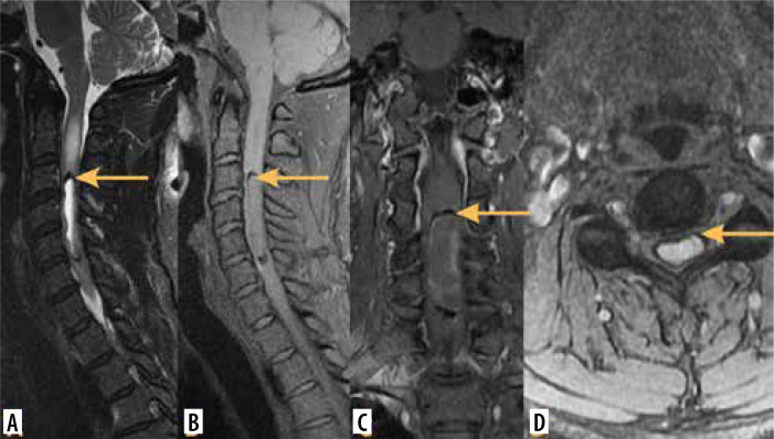
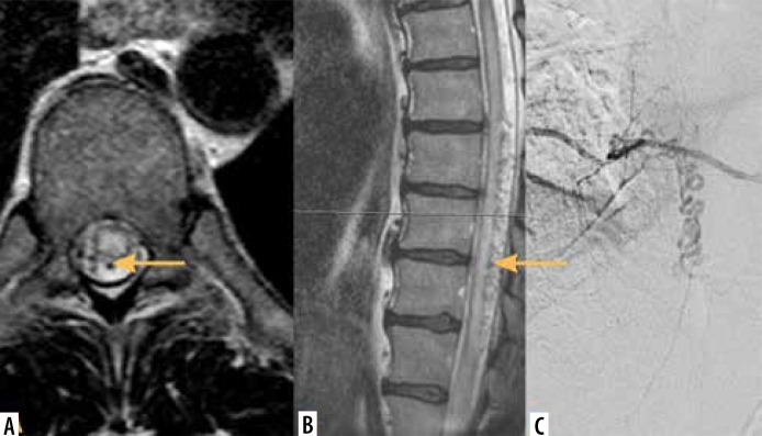
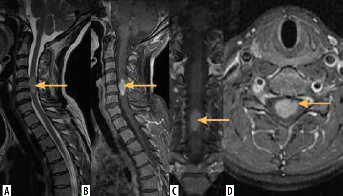
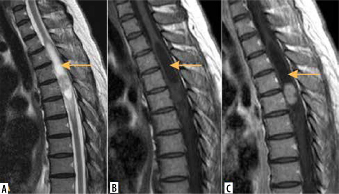
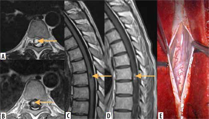
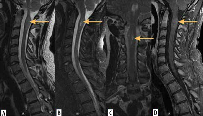
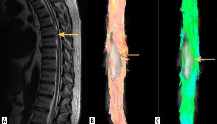
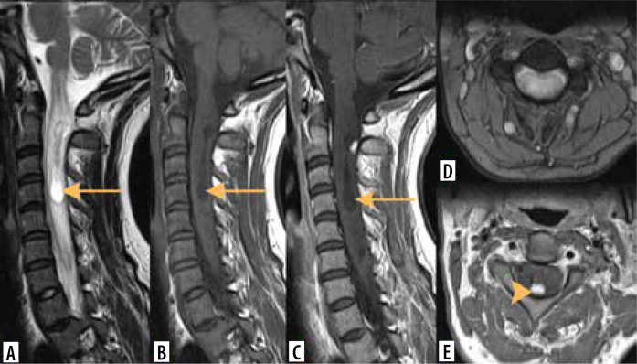
Intramedullary tumours (IMTs) are the least common neoplasms of the spinal canal. The majority of them are ependymomas and astrocytomas, the third commonest is haemangioblastoma, while other tumours of the spinal cord are relatively rare. This review presents on update on the imaging of spinal cord tumours. Magnetic resonance imaging (MRI) is the imaging method of choice in diagnosing IMTs, with other modalities playing a supplementary role. The authors discuss the MRI protocol in IMTs including advanced techniques and present the imaging features of particular tumours. The differentiation of IMTs from other spinal cord diseases is also presented.

摘要

髓内肿瘤(IMTs)是椎管内最不常见的肿瘤。其中大多数是室管膜瘤和星形细胞瘤,第三常见的是血管母细胞瘤,而脊髓的其他肿瘤相对少见。本综述介绍了脊髓肿瘤成像的最新情况。磁共振成像(MRI)是诊断IMTs的首选成像方法,其他方式起辅助作用。作者讨论了IMTs的MRI检查方案,包括先进技术,并介绍了特定肿瘤的成像特征。还介绍了IMTs与其他脊髓疾病的鉴别诊断。

https://cdn.ncbi.nlm.nih.gov/pmc/blobs/e193/11705043/96cd0a52c8ef/PJR-89-192424-g016.jpg
https://cdn.ncbi.nlm.nih.gov/pmc/blobs/e193/11705043/750f4fa2b19c/PJR-89-192424-g001.jpg
https://cdn.ncbi.nlm.nih.gov/pmc/blobs/e193/11705043/3aa191793c54/PJR-89-192424-g002.jpg
https://cdn.ncbi.nlm.nih.gov/pmc/blobs/e193/11705043/aa09cbadbaa5/PJR-89-192424-g003.jpg
https://cdn.ncbi.nlm.nih.gov/pmc/blobs/e193/11705043/b80bb57b15c3/PJR-89-192424-g004.jpg
https://cdn.ncbi.nlm.nih.gov/pmc/blobs/e193/11705043/d71e8c4859fb/PJR-89-192424-g005.jpg
https://cdn.ncbi.nlm.nih.gov/pmc/blobs/e193/11705043/253772e8a4a1/PJR-89-192424-g006.jpg
https://cdn.ncbi.nlm.nih.gov/pmc/blobs/e193/11705043/98a7b1053ddc/PJR-89-192424-g007.jpg
https://cdn.ncbi.nlm.nih.gov/pmc/blobs/e193/11705043/cc13468d12f3/PJR-89-192424-g008.jpg
https://cdn.ncbi.nlm.nih.gov/pmc/blobs/e193/11705043/c92550624a7b/PJR-89-192424-g009.jpg
https://cdn.ncbi.nlm.nih.gov/pmc/blobs/e193/11705043/830abff516c1/PJR-89-192424-g010.jpg
https://cdn.ncbi.nlm.nih.gov/pmc/blobs/e193/11705043/ad5e7428adc7/PJR-89-192424-g011.jpg
https://cdn.ncbi.nlm.nih.gov/pmc/blobs/e193/11705043/0468a7193cbe/PJR-89-192424-g012.jpg
https://cdn.ncbi.nlm.nih.gov/pmc/blobs/e193/11705043/def8f42f9f49/PJR-89-192424-g013.jpg
https://cdn.ncbi.nlm.nih.gov/pmc/blobs/e193/11705043/a7e580654270/PJR-89-192424-g014.jpg
https://cdn.ncbi.nlm.nih.gov/pmc/blobs/e193/11705043/077dfc880eb2/PJR-89-192424-g015.jpg
https://cdn.ncbi.nlm.nih.gov/pmc/blobs/e193/11705043/96cd0a52c8ef/PJR-89-192424-g016.jpg

相似文献

1
Imaging of intramedullary tumours of the spinal cord.脊髓髓内肿瘤的影像学检查
Pol J Radiol. 2024 Nov 15;89:e531-e540. doi: 10.5114/pjr/192424. eCollection 2024.
2
Magnetic Resonance Imaging Evaluation of Common Spinal Intramedullary Tumours: Ependymoma and Astrocytoma.磁共振成像评估常见脊髓髓内肿瘤:室管膜瘤和星形细胞瘤。
Mymensingh Med J. 2023 Jul;32(3):749-756.
3
Diagnosis of spinal cord ependymoma and astrocytic tumours with magnetic resonance imaging.脊髓室管膜瘤和星形细胞瘤的磁共振成像诊断
J Clin Neurosci. 1999 Mar;6(2):128-132. doi: 10.1054/jocn.1997.0005.
4
Intramedullary tumours and tumour mimics.髓内肿瘤和肿瘤样病变。
Clin Radiol. 2020 Nov;75(11):876.e17-876.e32. doi: 10.1016/j.crad.2020.05.010. Epub 2020 Jun 23.
5
Spinal perimedullary vein enlargement sign: an added value for the differentiation between intradural-extramedullary and intramedullary tumors on magnetic resonance imaging.脊髓髓周静脉增粗征:磁共振成像上区分硬脊膜内髓外肿瘤与髓内肿瘤的附加价值
Neuroradiology. 2016 Nov;58(11):1117-1124. doi: 10.1007/s00234-016-1744-4. Epub 2016 Sep 5.
6
[MRI symptomatology of primary intraspinal cord gliomas].[原发性脊髓内胶质瘤的磁共振成像症状学]
J Neuroradiol. 1995 Mar;22(1):28-42.
7
Microsurgical removal of intramedullary spinal cord tumours: report of 22 cases.显微手术切除脊髓髓内肿瘤:22例报告。
Acta Neurochir Suppl (Wien). 1988;43:9-12. doi: 10.1007/978-3-7091-8978-8_2.
8
[Intramedullary spinal cord tumors. Report. French Society of Neurosurgery. 45th annual congress. Angers, June 12-15 1994].[脊髓髓内肿瘤。报告。法国神经外科学会。第45届年会。昂热,1994年6月12日至15日]
Neurochirurgie. 1994;40 Suppl 1:1-108.
9
[Diagnosis of intramedullary spinal cord ependymomas and astrocytomas using magnetic resonance imaging: analysis of 15 cases].[利用磁共振成像诊断脊髓髓内室管膜瘤和星形细胞瘤:15例分析]
Di Yi Jun Yi Da Xue Xue Bao. 2004 Jul;24(7):847-8.
10
Exophytic ependymomas of the spinal cord.脊髓外生性室管膜瘤
Acta Neurochir (Wien). 2004 Sep;146(9):1047-50. doi: 10.1007/s00701-004-0275-7. Epub 2004 May 10.

本文引用的文献

1
Diagnosis of spinal dural arteriovenous fistula: a multimodal MRI assessment strategy.脊髓硬膜动静脉瘘的诊断:一种多模态 MRI 评估策略。
Clin Radiol. 2023 Dec;78(12):e958-e965. doi: 10.1016/j.crad.2023.08.027. Epub 2023 Sep 21.
2
Hemangioblastomas and Other Vascular Origating Tumors of Brain or Spinal Cord.脑或脊髓的血管母细胞瘤和其他血管起源性肿瘤。
Adv Exp Med Biol. 2023;1405:377-403. doi: 10.1007/978-3-031-23705-8_14.
3
Magnetic Resonance Imaging Evaluation of Common Spinal Intramedullary Tumours: Ependymoma and Astrocytoma.
磁共振成像评估常见脊髓髓内肿瘤:室管膜瘤和星形细胞瘤。
Mymensingh Med J. 2023 Jul;32(3):749-756.
4
Conventional and Advanced Imaging of Spinal Cord Tumors.脊髓肿瘤的常规与高级影像学表现。
Neuroimaging Clin N Am. 2023 Aug;33(3):389-406. doi: 10.1016/j.nic.2023.03.001. Epub 2023 May 18.
5
Surgical approaches to intramedullary spinal cord astrocytomas in the age of genomics.基因组学时代脊髓髓内星形细胞瘤的手术治疗方法
Front Oncol. 2022 Sep 6;12:982089. doi: 10.3389/fonc.2022.982089. eCollection 2022.
6
Spinal intramedullary hemangioblastoma and schwannoma collision tumor: illustrative case.脊髓髓内血管母细胞瘤与神经鞘瘤碰撞瘤:病例说明
J Neurosurg Case Lessons. 2021 Jan 18;1(3):CASE2059. doi: 10.3171/CASE2059.
7
[Spinal arteriovenous malformations].[脊髓动静脉畸形]
Radiologie (Heidelb). 2022 Aug;62(8):666-670. doi: 10.1007/s00117-022-01024-4. Epub 2022 Jun 29.
8
Neurofibromatosis from Head to Toe: What the Radiologist Needs to Know.从头至脚的神经纤维瘤病:放射科医生需要知道什么。
Radiographics. 2022 Jul-Aug;42(4):1123-1144. doi: 10.1148/rg.210235. Epub 2022 Jun 24.
9
MYXOPAPILLARY EPENDYMOMA OF THE SPINAL CORD IN ADULTS: A REPORT OF PERSONAL SERIES AND REVIEW OF LITERATURE.成人脊髓黏液乳头型室管膜瘤:个人系列报告和文献复习。
Acta Clin Croat. 2020 Jun;59(2):329-337. doi: 10.20471/acc.2020.59.02.17.
10
Syringomyelia and hydromyelia: Current understanding and neurosurgical management.脊髓空洞症和脑积水:当前的认识和神经外科治疗。
Rev Neurol (Paris). 2021 May;177(5):498-507. doi: 10.1016/j.neurol.2020.07.004. Epub 2020 Aug 18.