Evans Lowri E, Gray Anna L, Walsh Katy R, Danby Thea G E, Pritchard Harry A T, Allan Stuart M, Gurney Alison M, Greenstein Adam S, Schiessl Ingo
Geoffrey Jefferson Brain Research Centre, Manchester Academic Health Science Centre, Northern Care Alliance NHS Foundation Trust, University of Manchester, Manchester, UK.
Division of Cardiovascular Sciences, School of Biological Sciences, Faculty of Biology, Medicine and Health, University of Manchester, Manchester, UK.
Microcirculation. 2025 Jan;32(1):e70001. doi: 10.1111/micc.70001.
Cerebral blood flow (CBF) decline is increasingly recognized as an area of importance for targeting neurodegenerative disorders, yet full understanding of the mechanisms that underlie CBF changes are lacking. Animal models are crucial for expanding our knowledge as methods for studying global CBF and neurovascular coupling in humans are limited and require expensive specialized scanners.
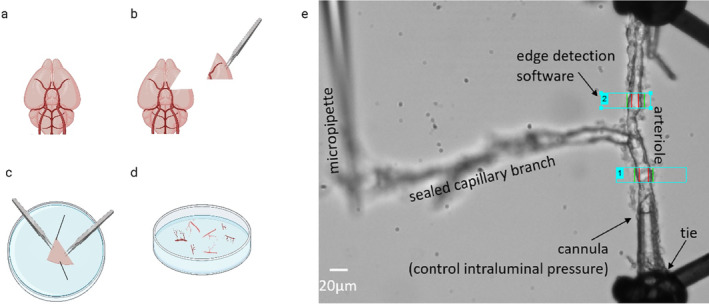
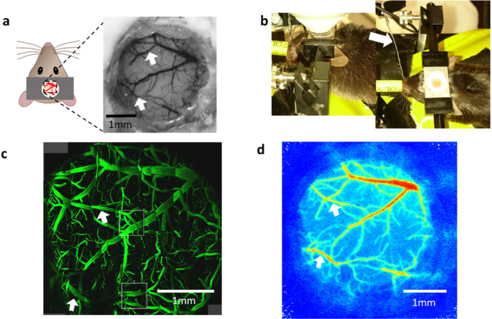
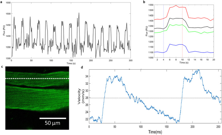
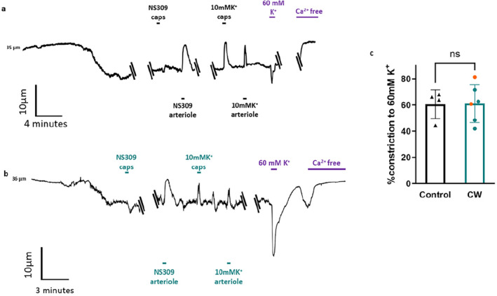
Use of appropriate animal models can increase our understanding of cerebrovascular function, so we have combined chronic cranial windows with in vivo two-photon and laser speckle microscopy and ex vivo capillary-parenchymal arteriole (CaPA) preparations. Chronic cranial windows allow for longitudinal direct observation of the cerebral microvasculature and surrounding parenchyma while the CaPA preparation can assess capillary and arteriole function in isolation of the neuronal tissue.
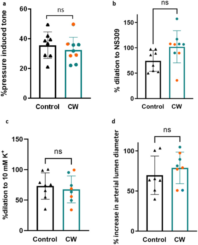
Here, we found that extra-dural cranial windows and related imaging protocols do not affect vascular function in the CaPA preparation. Cortical vessels from animals that have undergone imaging can therefore be taken to discover physiological alterations in the cerebral vasculature that contribute to any observed in vivo changes.
This approach will enhance neurodegenerative research with the benefit of limiting animal usage.
脑血流量(CBF)下降日益被视为针对神经退行性疾病的一个重要领域,但对CBF变化背后的机制仍缺乏全面了解。动物模型对于扩展我们的知识至关重要,因为研究人类全脑CBF和神经血管耦合的方法有限,且需要昂贵的专业扫描仪。
使用合适的动物模型可以增进我们对脑血管功能的理解,因此我们将慢性颅骨视窗与体内双光子和激光散斑显微镜以及体外毛细血管 - 实质小动脉(CaPA)制备相结合。慢性颅骨视窗允许对脑微血管和周围实质进行纵向直接观察,而CaPA制备可以在分离神经元组织的情况下评估毛细血管和小动脉的功能。
在此,我们发现硬脑膜外颅骨视窗及相关成像方案不会影响CaPA制备中的血管功能。因此,可以取用经过成像的动物的皮质血管来发现脑脉管系统中的生理改变,这些改变导致了任何观察到的体内变化。
这种方法将通过限制动物使用量来加强神经退行性疾病的研究。