Marquart Grant W, Stoddard Jonathan, Kinnison Karen, Zhou Felicia, Hugo Richard, Ryals Renee, Shubert Scott, McGill Trevor J, Mackiewicz Marilyn R
Department of Chemistry, Portland State University, Portland, Oregon 97207, United States.
Casey Eye Institute, Oregon Health & Science University, Portland, Oregon 97239, United States.
ACS Appl Nano Mater. 2022 May 27;5(5):6995-7008. doi: 10.1021/acsanm.2c00958. Epub 2022 May 16.
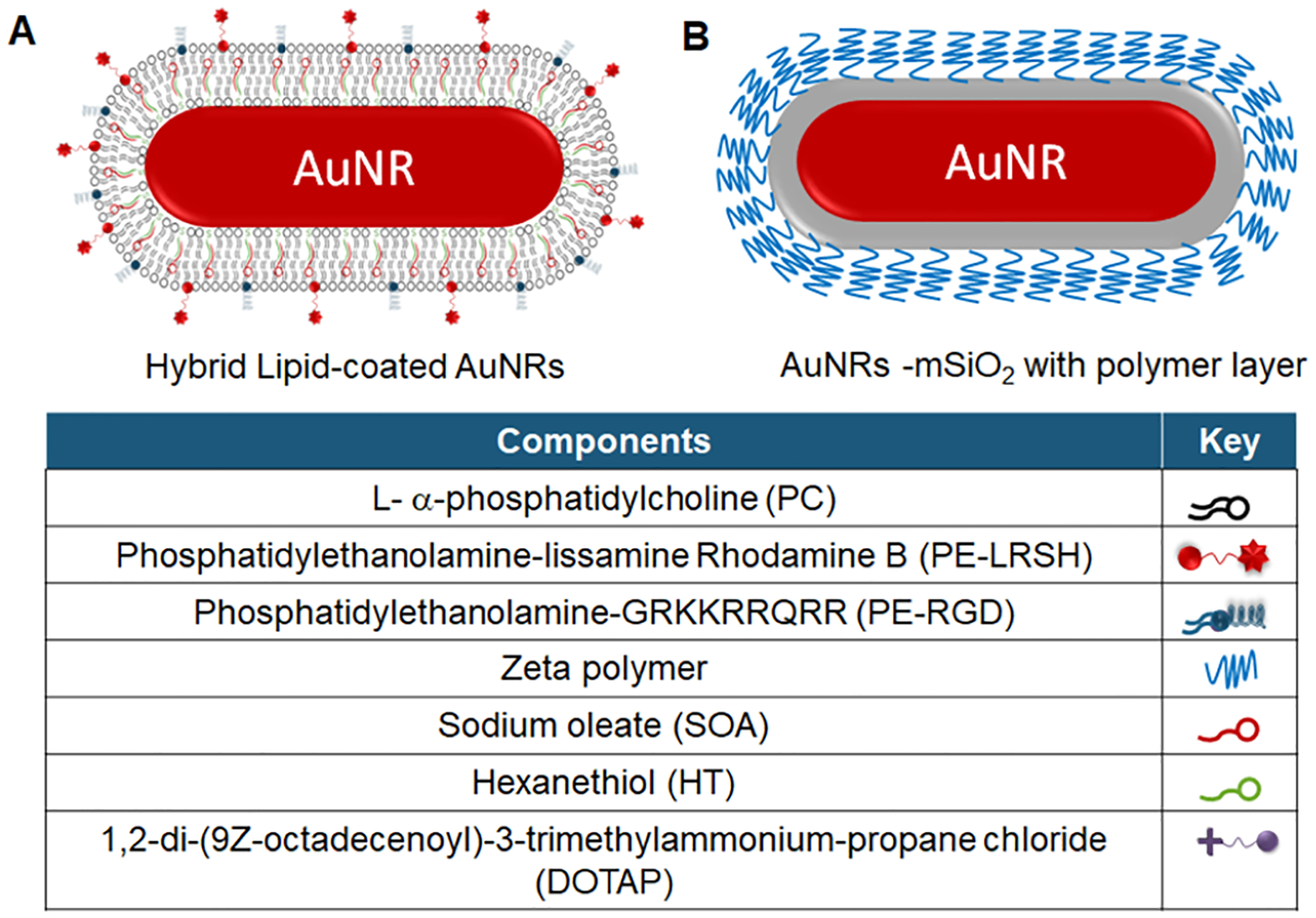
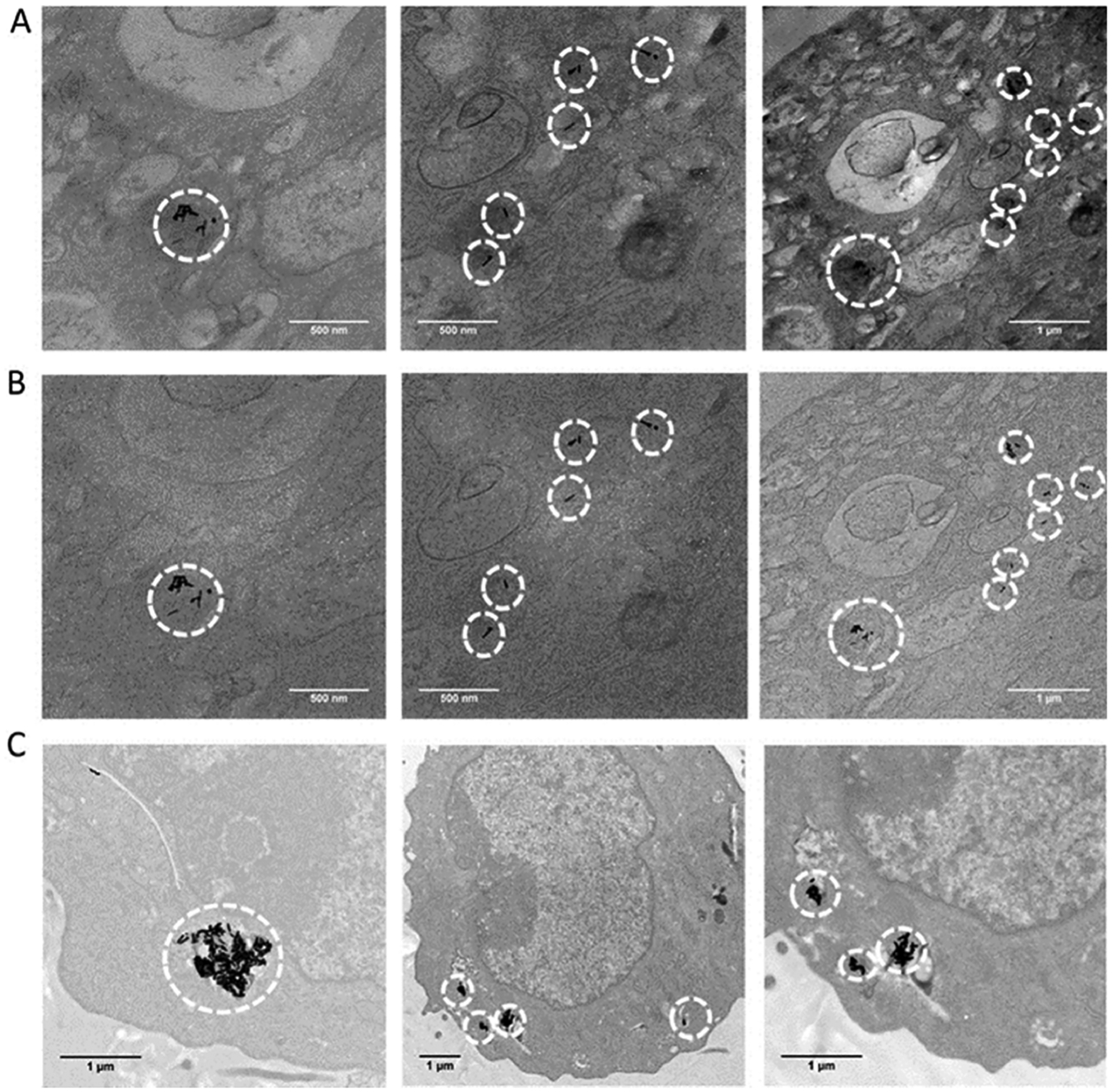
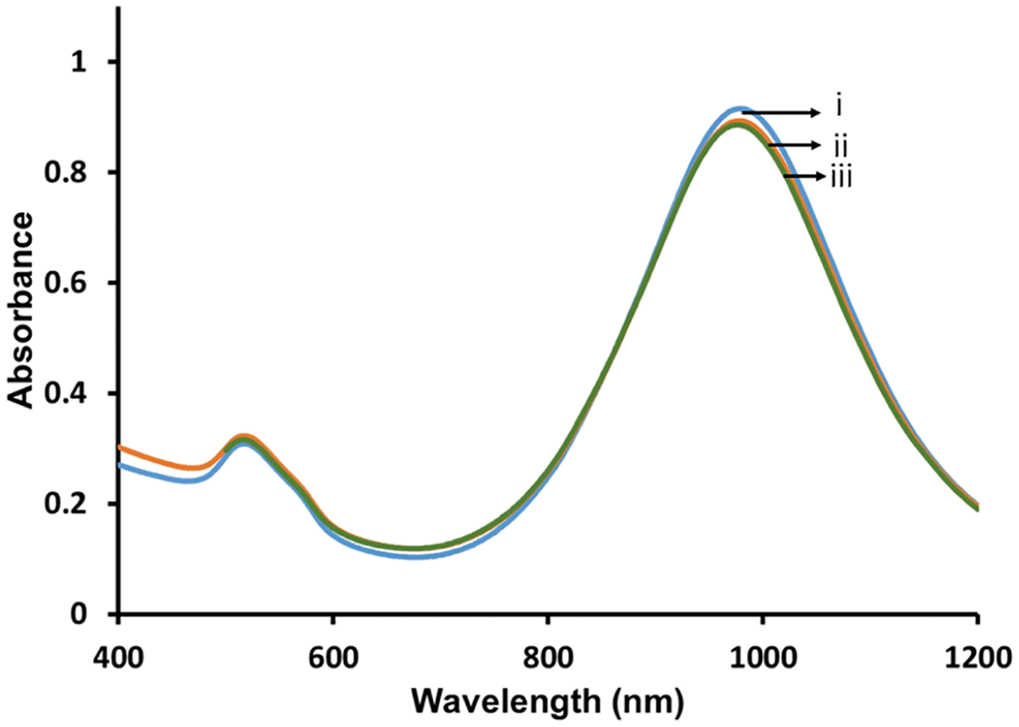
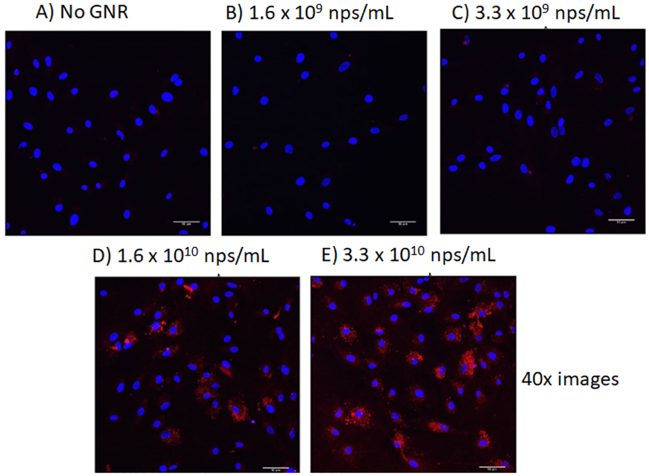
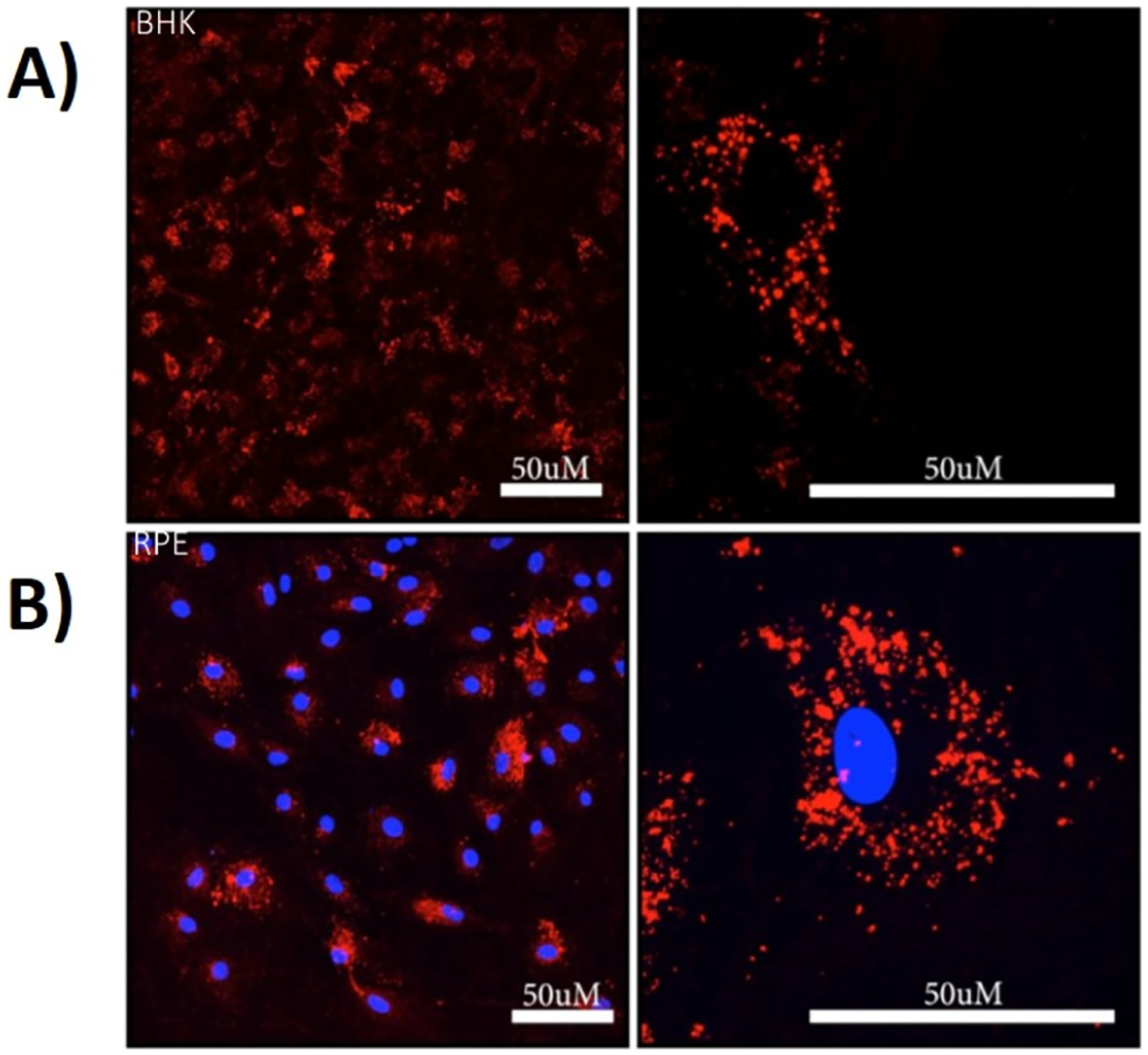
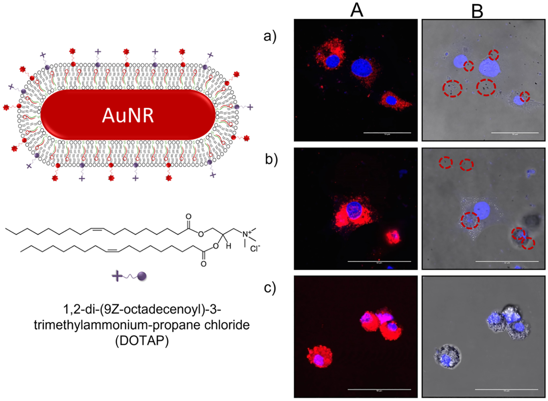
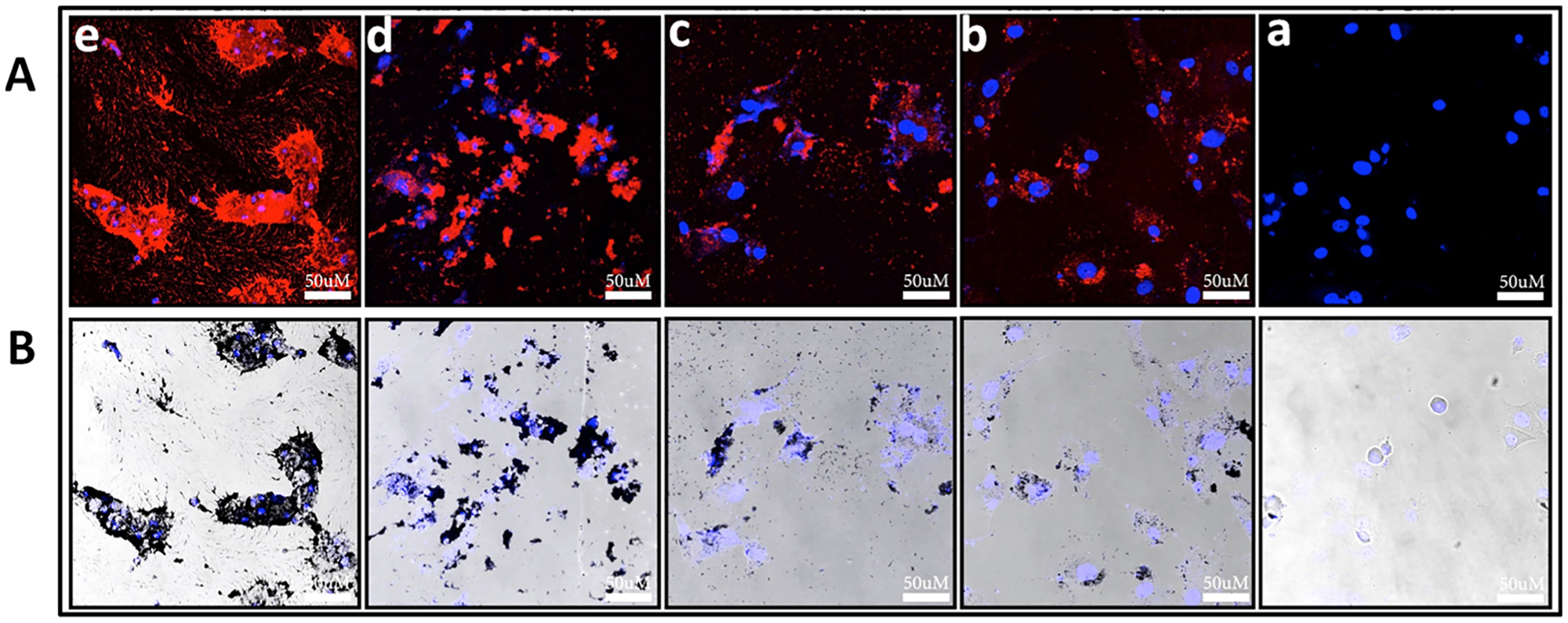
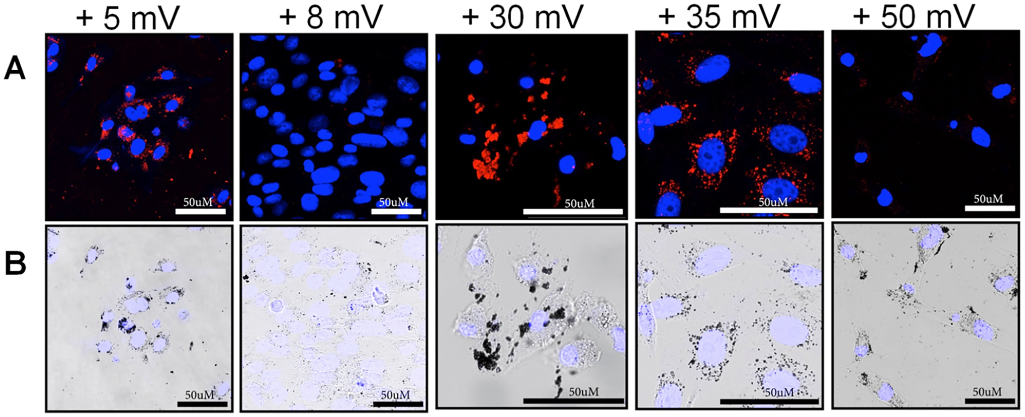
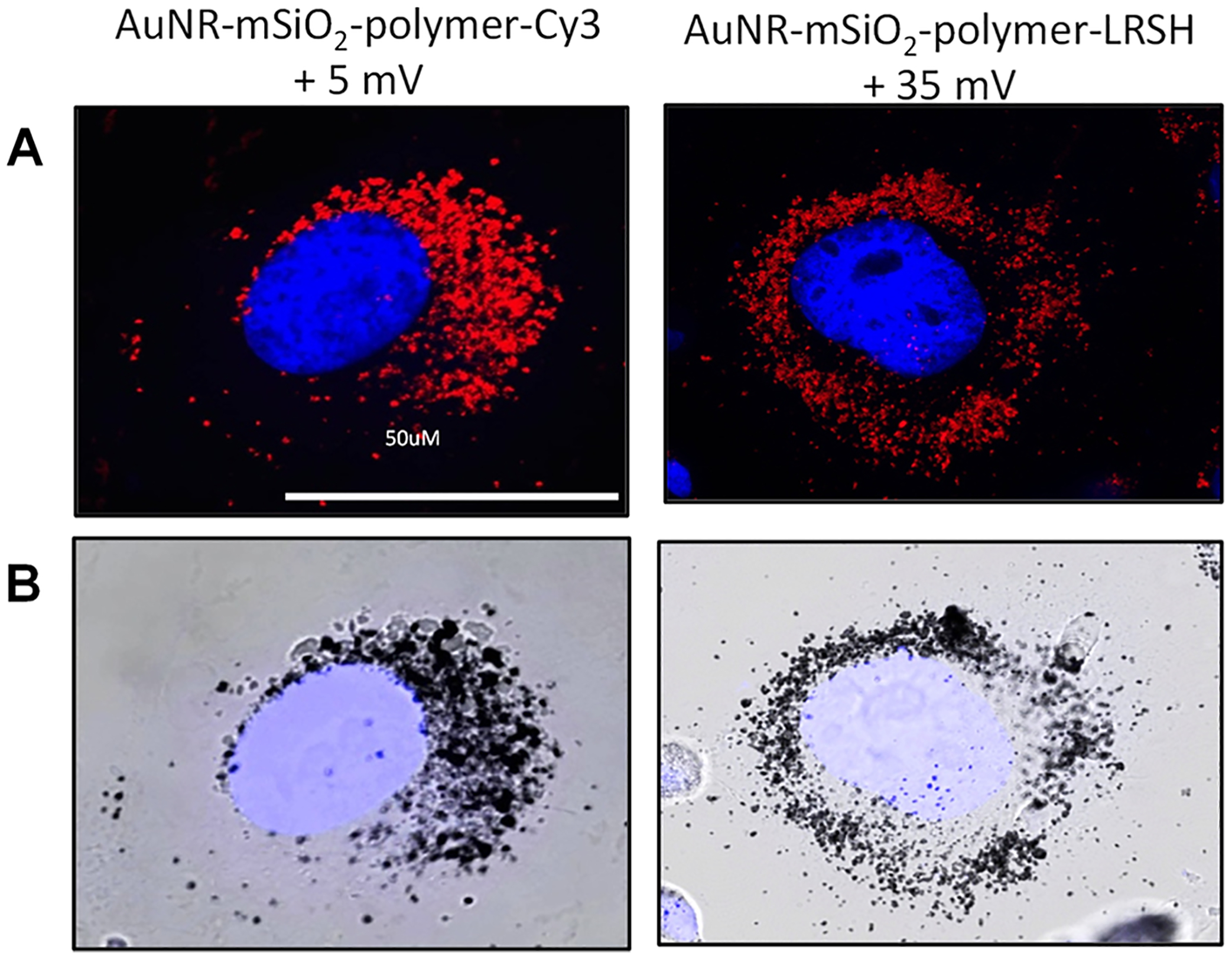
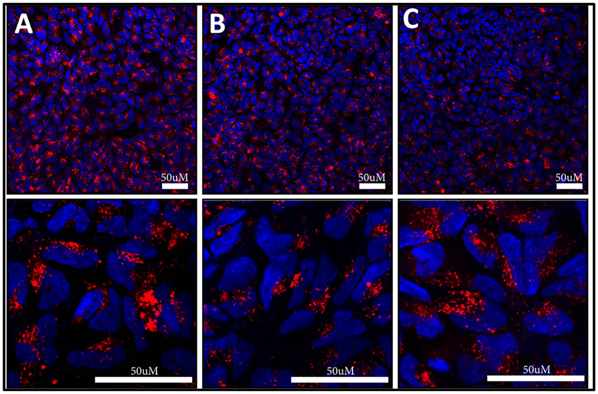
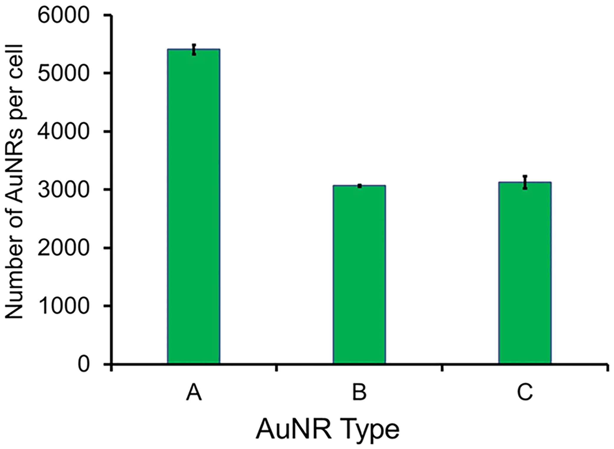
The advancement of safe nanomaterials for use as optical coherence tomography (OCT) imaging and stem cell-labeling agents to longitudinally visually track therapeutic derived retinal stem cells to study their migration, survival rate, and efficacy is challenged by instability, intracellular aggregation, low uptake, and cytotoxicity. Here, we describe a series of hybrid lipid-coated gold nanorods (AuNRs) that could solve these issues. These nanomaterials were made via a layer-by-layer assembly approach, and their stability in biological media, mechanism, efficiency of uptake, and toxicity were compared with a commercially available set of AuNRs with a 5 nm mesoporous silica (mSiO)-polymer coating. These nanomaterials can serve as stem cell labeling and OCT imaging agents because they absorb in the near-infrared (NIR) region away from biological tissues. Although both subtypes of AuNRs were taken up by retinal pigment epithelial, neural progenitor, and baby hamster kidney cells, slightly negatively charged hybrid lipid-coated AuNRs had minimal aggregation in biological media and within the cytoplasm of cells (3000 AuNRs/cell) as well as minimal impact on cell health. Hybrid lipid-coated AuNRs modified with cell-penetrating peptides had the least toxicological impact, with >92% cell viability. In contrast, the more "sticky" AuNRs with a 5 nm mSiO-polymer coating showed significant aggregation in biological media and within the cytoplasm with lower-than-expected uptake of AuNRs (5400 of AuNRs/cell) given their highly positive surface charge (35+ mV). Collectively, we have demonstrated that hybrid lipid-coated AuNRs, which absorb in the NIR-II region away from biological tissues, with tuned surface chemistry can label therapeutic derived stem cells with minimal aggregation and impact on cell health as well as enhance uptake for OCT imaging applications.
将安全的纳米材料用作光学相干断层扫描(OCT)成像和干细胞标记剂,以纵向可视化追踪治疗性视网膜干细胞,研究其迁移、存活率和疗效,这受到不稳定性、细胞内聚集、低摄取率和细胞毒性的挑战。在此,我们描述了一系列能够解决这些问题的杂化脂质包覆金纳米棒(AuNRs)。这些纳米材料是通过逐层组装方法制备的,并将其在生物介质中的稳定性、作用机制、摄取效率和毒性与一组市售的带有5纳米介孔二氧化硅(mSiO)-聚合物涂层的AuNRs进行了比较。这些纳米材料可作为干细胞标记和OCT成像剂,因为它们在远离生物组织的近红外(NIR)区域有吸收。尽管两种亚型的AuNRs都被视网膜色素上皮细胞、神经祖细胞和幼仓鼠肾细胞摄取,但带轻微负电荷的杂化脂质包覆AuNRs在生物介质和细胞胞质内的聚集最小(约3000个AuNRs/细胞),对细胞健康的影响也最小。用细胞穿透肽修饰的杂化脂质包覆AuNRs的毒理学影响最小,细胞活力>92%。相比之下,带有5纳米mSiO-聚合物涂层的更“黏附性”的AuNRs在生物介质和胞质内显示出显著聚集,鉴于其高度正表面电荷(35+ mV),其AuNRs摄取率低于预期(约5400个AuNRs/细胞)。总体而言,我们已经证明,在远离生物组织的近红外II区域有吸收、具有可调表面化学性质的杂化脂质包覆AuNRs能够以最小的聚集和对细胞健康的影响标记治疗性干细胞,并提高摄取率以用于OCT成像应用。