Hameed Shahna S, Sharma Tasneem P
Department of Ophthalmology, Indiana University School of Medicine, Indianapolis, Indiana.
Curr Protoc. 2025 Jan;5(1):e70091. doi: 10.1002/cpz1.70091.
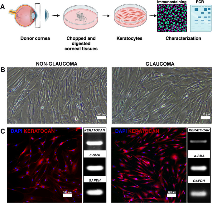
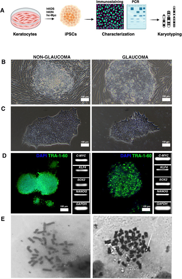
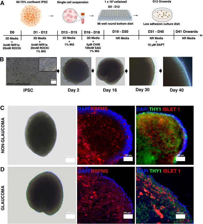
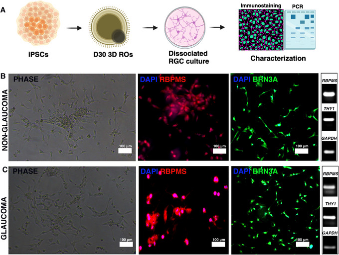
Human induced pluripotent stem cell (hiPSC)-based disease modeling can be successfully recapitulated to mimic disease characteristics across various human pathologies. Glaucoma, a progressive optic neuropathy, primarily affects the retinal ganglion cells (RGCs). While multiple groups have successfully generated RGCs from non-diseased hiPSCs, producing RGCs from glaucomatous human samples holds significant promise for understanding disease pathology by revealing patient-specific disease signatures. Given that keratocytes originate from the neural crest and previous reports suggest that ocular fibroblasts from glaucomatous donors carry pathogenic signatures, it is highly plausible that these signatures imprinted within the keratocytes will also be present in the derived RGCs. Thus, we aimed to generate RGCs from both glaucomatous and non-glaucomatous donor keratocytes and validate disease-specific signatures in 3D retinal organoids and in isolated RGCs. Our protocol describes the generation of iPSCs from keratocytes of both glaucomatous and non-glaucomatous donors, followed by their differentiation into retinal organoids. Subsequent isolation and culturing of RGCs were performed. Disease signatures in the RGCs were validated in both 3D retinal organoids (ROs) and 2D RGC cultures, and glaucomatous RGCs in 3D and 2D cultures demonstrated increased cleaved CASP3 and significant RGC loss, indicating disease imprints in the hiPSC-derived RGCs. This model offers a venue and high throughput platform for studying glaucomatous disease pathology and holds significant potential for drug discovery using RGCs derived from human donors. © 2025 The Author(s). Current Protocols published by Wiley Periodicals LLC. Basic Protocol 1: Culturing of keratocytes from human cadaveric donors Basic Protocol 2: Reprogramming donor keratocytes into iPSCs Basic Protocol 3: Evaluation of chromosomal loss during reprogramming in iPSCs by karyotyping Basic Protocol 4: Generation of 3D ROs Basic Protocol 5: Dissociation and culturing of RGCs from 3D ROs Support Protocol 1: Immunostaining for phenotypic characterization of cells Support Protocol 2: Sectioning of 3D ROs and immunostaining Support Protocol 3: Western blotting for cleaved CASP3 and THY1.
基于人诱导多能干细胞(hiPSC)的疾病建模能够成功地模拟各种人类病理状况下的疾病特征。青光眼是一种进行性视神经病变,主要影响视网膜神经节细胞(RGCs)。虽然多个研究小组已成功从非患病的hiPSC中生成RGCs,但从青光眼患者样本中生成RGCs对于通过揭示患者特异性疾病特征来理解疾病病理具有重要意义。鉴于角膜细胞起源于神经嵴,且先前的报告表明青光眼供体的眼成纤维细胞携带致病特征,极有可能在角膜细胞中印刻的这些特征也会存在于所衍生的RGCs中。因此,我们旨在从青光眼和非青光眼供体的角膜细胞中生成RGCs,并在3D视网膜类器官和分离的RGCs中验证疾病特异性特征。我们的方案描述了从青光眼和非青光眼供体的角膜细胞中生成iPSC,随后将其分化为视网膜类器官。接着进行RGCs的后续分离和培养。在3D视网膜类器官(ROs)和2D RGC培养物中均验证了RGCs中的疾病特征,并且3D和2D培养物中的青光眼RGCs显示出裂解的CASP3增加以及RGCs显著损失,表明在hiPSC衍生的RGCs中存在疾病印记。该模型为研究青光眼疾病病理提供了一个场所和高通量平台,并且在使用源自人类供体的RGCs进行药物发现方面具有巨大潜力。© 2025作者。由Wiley Periodicals LLC出版的《当前方案》。基本方案1:从人类尸体供体培养角膜细胞 基本方案2:将供体角膜细胞重编程为iPSC 基本方案3:通过核型分析评估iPSC重编程过程中的染色体丢失 基本方案4:生成3D ROs 基本方案5:从3D ROs中解离并培养RGCs 支持方案1:用于细胞表型特征的免疫染色 支持方案2:3D ROs切片及免疫染色 支持方案3:针对裂解的CASP3和THY1的蛋白质免疫印迹法