Boal Andrew M, McGrady Nolan R, Holden Joseph M, Risner Michael L, Calkins David J
Department of Ophthalmology and Visual Sciences, Vanderbilt Eye Institute, Vanderbilt University Medical Center, Nashville, TN, United States.
Front Neurosci. 2023 Mar 27;17:1142668. doi: 10.3389/fnins.2023.1142668. eCollection 2023.
Identification of early adaptive and maladaptive neuronal stress responses is an important step in developing targeted neuroprotective therapies for degenerative disease. In glaucoma, retinal ganglion cells (RGCs) and their axons undergo progressive degeneration resulting from stress driven by sensitivity to intraocular pressure (IOP). Despite therapies that can effectively manage IOP many patients progress to vision loss, necessitating development of neuronal-based therapies. Evidence from experimental models of glaucoma indicates that early in the disease RGCs experience altered excitability and are challenged with dysregulated potassium (K) homeostasis. Previously we demonstrated that certain RGC types have distinct excitability profiles and thresholds for depolarization block, which are associated with sensitivity to extracellular K.
Here, we used our inducible mouse model of glaucoma to investigate how RGC sensitivity to K changes with exposure to elevated IOP.
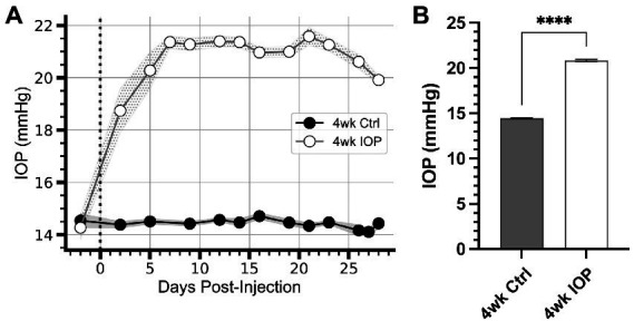
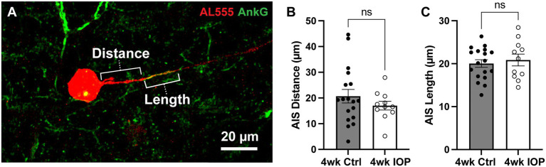
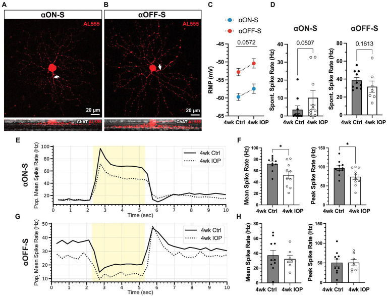
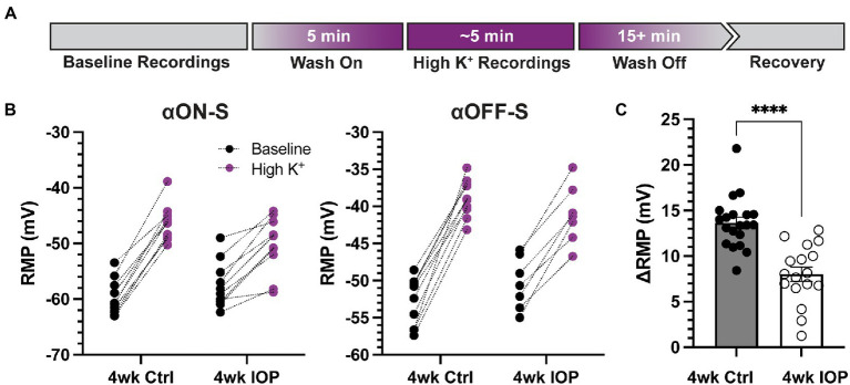
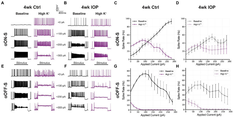
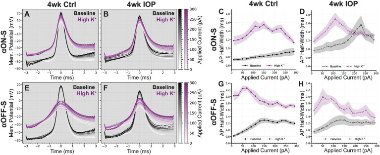
In controls, conditions of increased K enhanced membrane depolarization, reduced action potential generation, and widened action potentials. Consistent with our previous work, 4 weeks of IOP elevation diminished RGC light-and current-evoked responses. Compared to controls, we found that IOP elevation reduced the effects of increased K on depolarization block threshold, with IOP-exposed cells maintaining greater excitability. Finally, IOP elevation did not alter axon initial segment dimensions, suggesting that structural plasticity alone cannot explain decreased K sensitivity.
Thus, in response to prolonged IOP elevation RGCs undergo an adaptive process that reduces sensitivity to changes in K while diminishing excitability. These experiments give insight into the RGC response to IOP stress and lay the groundwork for mechanistic investigation into targets for neuroprotective therapy.
识别早期适应性和适应不良的神经元应激反应是开发针对退行性疾病的靶向神经保护疗法的重要一步。在青光眼患者中,视网膜神经节细胞(RGCs)及其轴突会因眼内压(IOP)敏感性所驱动的应激而逐渐退化。尽管有能有效控制眼压的疗法,但许多患者仍会进展至视力丧失,因此需要开发基于神经元的疗法。青光眼实验模型的证据表明,在疾病早期,RGCs的兴奋性会发生改变,并面临钾(K)稳态失调的挑战。此前我们证明,某些RGC类型具有不同的兴奋性特征和去极化阻滞阈值,这与对细胞外K的敏感性有关。
在此,我们使用可诱导的青光眼小鼠模型来研究RGCs对K的敏感性如何随眼压升高而变化。
在对照组中,K增加的情况下会增强膜去极化、减少动作电位的产生并使动作电位变宽。与我们之前的研究一致,眼压升高4周会减弱RGCs的光诱发反应和电流诱发反应。与对照组相比,我们发现眼压升高会降低K增加对去极化阻滞阈值的影响,眼压升高组的细胞保持更高的兴奋性。最后,眼压升高并未改变轴突起始段的尺寸,这表明仅结构可塑性无法解释K敏感性降低的原因。
因此,对长期眼压升高的反应中,RGCs会经历一个适应性过程,该过程会降低对K变化的敏感性,同时降低兴奋性。这些实验深入了解了RGCs对眼压应激的反应,并为神经保护治疗靶点的机制研究奠定了基础。