Oostenbrink Akkelien H A, Bronkhorst Ewald M, Booij Johan W, Dieters Arjan J A, Ren Yijin, Kuijpers-Jagtman Anne Marie, Bruggink Robin
Department of Orthodontics, University Medical Center Groningen, University of Groningen, Hanzeplein 1, 9713 GZ Groningen, The Netherlands.
Department of Dentistry, Radboud Research Institute for Medical Innovation, Radboud University Medical Center, Philips van Leijdenlaan 25, 6525 EX Nijmegen, The Netherlands.
J Clin Med. 2025 Jan 3;14(1):225. doi: 10.3390/jcm14010225.
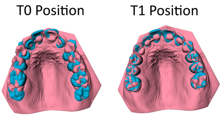
This retrospective longitudinal outcome study comparing orthodontic extraction modalities, including extraction of maxillary first or second molars, aimed to compare the three-dimensional tooth movement of maxillary canines (C), premolars (P1, P2), and molars (M1, M2) in Class II division 1 malocclusion treatment with fixed appliances. A sample of 98 patients (mean age 13.20 ± 1.46 years) was selected for the M1 group, and 64 patients (mean age 13.20 ± 1.36 years) were chosen for the M2 group. Tooth movement was analyzed three-dimensionally on pre-treatment (T0) and post-treatment (T1) digital dental casts. Regression analyses compared the tooth movements (in mm) between the M1 and M2 groups. The mean treatment duration for the M1 group was 2.51 ± 0.55 year, while, for the M2 group, it was 1.53 ± 0.37 year. The data showed limited distal movements of the C, P1, and P2 of approximately 2 mm in the M1 group and 1 mm in the M2 group during orthodontic treatment, but the M1 group exhibited significantly more distal movements than the M2 group (mean difference 1.11 to 1.24 mm). Vertical movements of the C, P1, and P2 in both groups were also minor (0.16 to 1.26 mm). The differences between groups did not exceed 0.2 mm and were not significant. Both treatment modalities resulted in a significant degree of anchorage loss with a distinct mesialization (8.40 ± 1.66 mm) of M2 in the M1 group and limited distalization (0.83 ± 0.98 mm) of M1 in the M2 group. The findings highlight the importance of thorough case evaluation when choosing between extraction modalities in Class II treatment. If a large distal movement of canines and premolars is required, additional anchorage mechanics should be considered.
这项回顾性纵向结果研究比较了正畸拔牙方式,包括拔除上颌第一或第二磨牙,旨在比较在安氏II类1分类错 畸形固定矫治中上颌尖牙(C)、前磨牙(P1、P2)和磨牙(M1、M2)的三维牙齿移动情况。M1组选取了98例患者(平均年龄13.20±1.46岁),M2组选取了64例患者(平均年龄13.20±1.36岁)。在治疗前(T0)和治疗后(T1)的数字化牙模上对牙齿移动进行三维分析。回归分析比较了M1组和M2组之间的牙齿移动(以毫米为单位)。M1组的平均治疗时间为2.51±0.55年,而M2组为1.53±0.37年。数据显示,在正畸治疗期间,M1组的C、P1和P2远中移动有限,约为2毫米,M2组为1毫米,但M1组的远中移动明显多于M2组(平均差异为1.11至1.24毫米)。两组中C、P1和P2的垂直移动也较小(0.16至1.26毫米)。组间差异不超过0.2毫米,无统计学意义。两种治疗方式均导致明显的支抗丧失,M1组中M2有明显的近中移动(8.40±1.66毫米),M2组中M1有有限的远中移动(0.83±0.98毫米)。研究结果突出了在II类治疗中选择拔牙方式时进行全面病例评估的重要性。如果需要尖牙和前磨牙进行较大的远中移动,则应考虑额外的支抗机制。