Stellpflug Austin, Caron Justin, Fasciano Samantha, Wang Bo, Wang Shue
Joint Department of Biomedical Engineering, Marquette University and the Medical College of Wisconsin Milwaukee WI 53226 USA
Department of Chemistry, Chemical and Biomedical Engineering, University of New Haven West Haven CT 06516 USA
Nanoscale Adv. 2024 Dec 27;7(3):735-747. doi: 10.1039/d4na00797b. eCollection 2025 Jan 28.
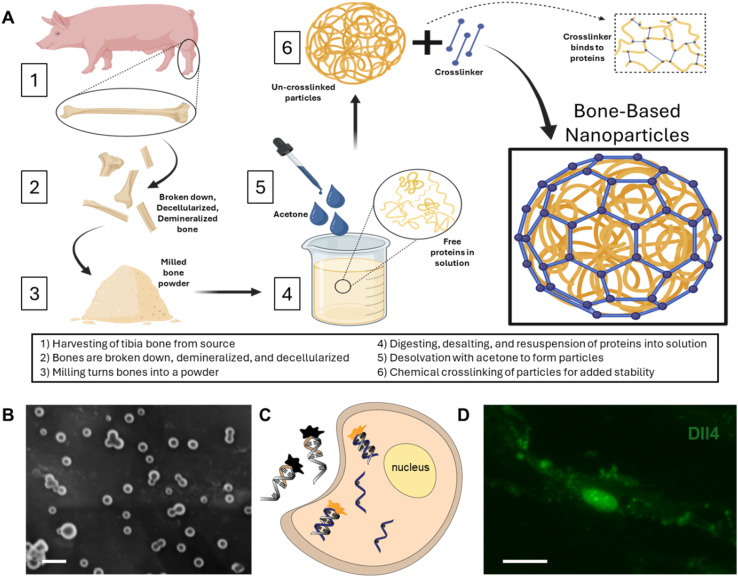
Mesenchymal stem cell (MSC)-based bone tissue regeneration has gained significant attention due to the excellent differentiation capacity and immunomodulatory activity of MSCs. Enhancing osteogenesis regulation is crucial for improving the therapeutic efficacy of MSC-based regeneration. By utilizing the regenerative capacity of bone ECM and the functionality of nanoparticles, we recently engineered bone-based nanoparticles (BNPs) from decellularized porcine bones. The effects of internalization of BNPs on MSC viability, proliferation, and osteogenic differentiation were first investigated and compared at different time points. The phenotypic behaviors, including cell number, proliferation, and differentiation were characterized and compared. By incorporating a LNA/DNA nanobiosensor and MSC live cell imaging, we monitored and compared Notch ligand delta-like 4 (Dll4) expression dynamics in the cytoplasm and nucleus during osteogenic differentiation. Pharmacological interventions are used to inhibit Notch signaling to examine the mechanisms involved. The results suggest that Notch inhibition mediates the osteogenic process, with reduced expression of early and late stage differentiation markers (ALP and calcium mineralization). The internalization of BNPs led to an increase in Dll4 expression, exhibiting a time-dependent pattern that aligned with enhanced cell proliferation and differentiation. Our findings indicate that the observed changes in BNP-treated cells during osteogenic differentiation could be associated with elevated levels of Dll4 mRNA expression. In summary, this study provides new insights into MSC osteogenic differentiation and the molecular mechanisms through which BNPs stimulate this process. The results indicate that BNPs influence osteogenesis by modulating Notch ligand Dll4 expression, demonstrating a potential link between Notch signaling and the proteins present in BNPs.
基于间充质干细胞(MSC)的骨组织再生因其出色的分化能力和免疫调节活性而备受关注。增强成骨调节对于提高基于MSC的再生治疗效果至关重要。通过利用骨细胞外基质(ECM)的再生能力和纳米颗粒的功能,我们最近从脱细胞猪骨中构建了骨基纳米颗粒(BNP)。首先在不同时间点研究并比较了BNP内化对MSC活力、增殖和成骨分化的影响。对包括细胞数量、增殖和分化在内的表型行为进行了表征和比较。通过整合锁核酸/DNA纳米生物传感器和MSC活细胞成像,我们监测并比较了成骨分化过程中细胞质和细胞核中Notch配体Delta样4(Dll4)的表达动态。使用药物干预抑制Notch信号传导以研究其中涉及的机制。结果表明,Notch抑制介导了成骨过程,早期和晚期分化标志物(碱性磷酸酶和钙矿化)的表达降低。BNP的内化导致Dll4表达增加,呈现出与细胞增殖和分化增强相一致的时间依赖性模式。我们的研究结果表明,在成骨分化过程中BNP处理的细胞中观察到的变化可能与Dll4 mRNA表达水平升高有关。总之,本研究为MSC成骨分化以及BNP刺激这一过程的分子机制提供了新的见解。结果表明,BNP通过调节Notch配体Dll4的表达影响成骨,证明了Notch信号传导与BNP中存在的蛋白质之间的潜在联系。