Zhang Li, Wen Xin, Ma Jing-Wen, Wang Jian-Wei, Huang Yao, Wu Ning, Li Meng
Department of Diagnostic Radiology, National Cancer Center/National Clinical Research Center for Cancer/Cancer Hospital, Chinese Academy of Medical Sciences and Peking Union Medical College, Beijing, China.
Department of Radiology, State Key Laboratory of Cardiovascular Disease, National Clinical Research Center for Cardiovascular Diseases, Fuwai Hospital, Chinese Academy of Medical Sciences and Peking Union Medical College, Beijing, China.
J Thorac Dis. 2024 Dec 31;16(12):8782-8795. doi: 10.21037/jtd-24-1125. Epub 2024 Dec 27.
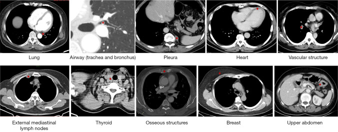
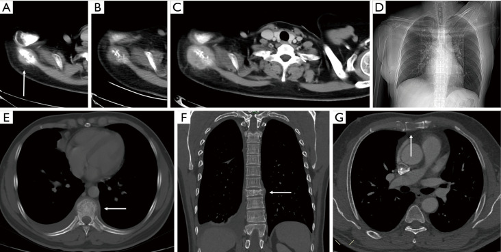
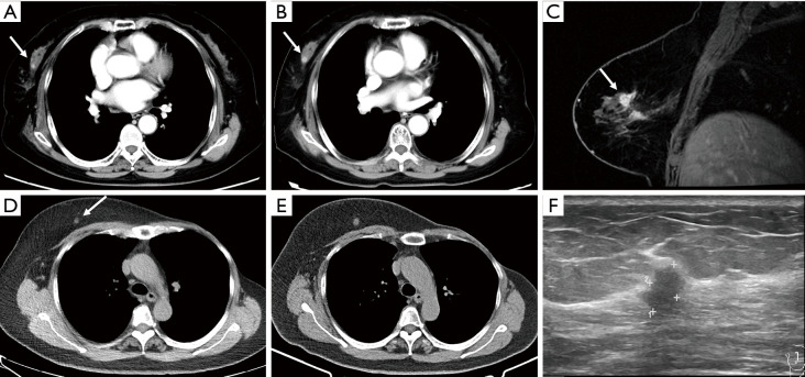
Chest computed tomography (CT) is the most frequently performed imaging examination worldwide. Compared with chest radiography, chest CT greatly improves the detection rate and diagnostic accuracy of chest lesions because of the absence of overlapping structures and is the best imaging technique for the observation of chest lesions. However, there are still frequently missed diagnoses during the interpretation process, especially in certain areas or "blind spots", which may possibly be overlooked by radiologists. Awareness of these blind spots is of great significance to avoid false negative results and potential adverse consequences for patients. In this review, we summarize the common blind spots identified in actual clinical practice, encompassing the central areas within the pulmonary parenchyma (including the perihilar regions, paramediastinal regions, and operative area after surgery), trachea and bronchus, pleura, heart, vascular structure, external mediastinal lymph nodes, thyroid, osseous structures, breast, and upper abdomen. In addition to careful review, clinicians can employ several techniques to mitigate or minimize errors arising from these blind spots in film interpretation and reporting. In this review, we also propose technical methods to reduce missed diagnoses, including advanced imaging post-processing techniques such as multiplanar reconstruction (MPR), maximum intensity projection (MIP), artificial intelligence (AI) and structured reporting which can significantly enhance the detection of lesions and improve diagnostic accuracy.
胸部计算机断层扫描(CT)是全球最常进行的影像学检查。与胸部X线摄影相比,胸部CT由于不存在结构重叠,大大提高了胸部病变的检出率和诊断准确性,是观察胸部病变的最佳影像学技术。然而,在解读过程中仍经常出现漏诊情况,尤其是在某些区域或“盲点”,放射科医生可能会忽略这些区域。了解这些盲点对于避免假阴性结果以及对患者可能产生的不良后果具有重要意义。在本综述中,我们总结了在实际临床实践中发现的常见盲点,包括肺实质内的中央区域(包括肺门周围区域、纵隔旁区域和术后手术区域)、气管和支气管、胸膜、心脏、血管结构、纵隔外淋巴结、甲状腺、骨骼结构、乳房和上腹部。除了仔细阅片外,临床医生可以采用多种技术来减轻或尽量减少在胶片解读和报告中因这些盲点而产生的错误。在本综述中,我们还提出了减少漏诊的技术方法,包括先进的影像后处理技术,如多平面重建(MPR)、最大密度投影(MIP)、人工智能(AI)和结构化报告,这些技术可以显著提高病变的检出率并提高诊断准确性。