Department of Radiology, Medical University of South Carolina, Charleston, SC, 29403, USA.
Siemens Healthineers, Princeton, NJ, USA.
BMC Med. 2021 Mar 4;19(1):55. doi: 10.1186/s12916-021-01928-3.
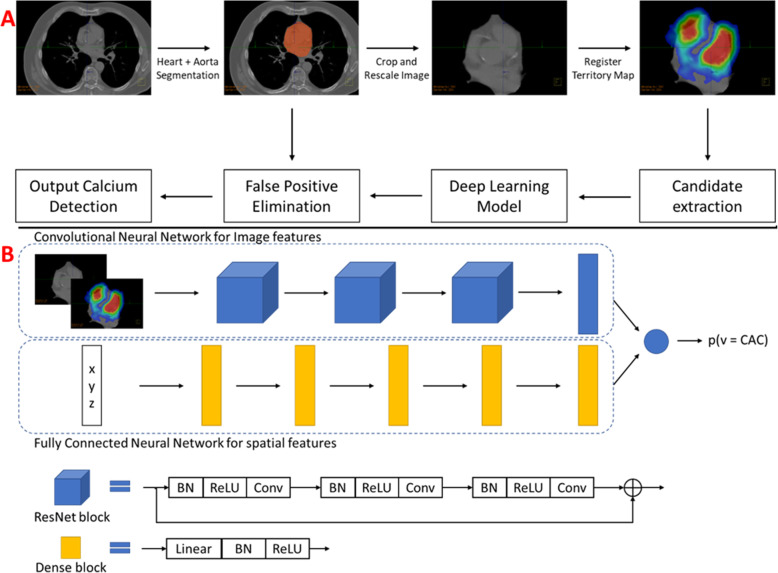
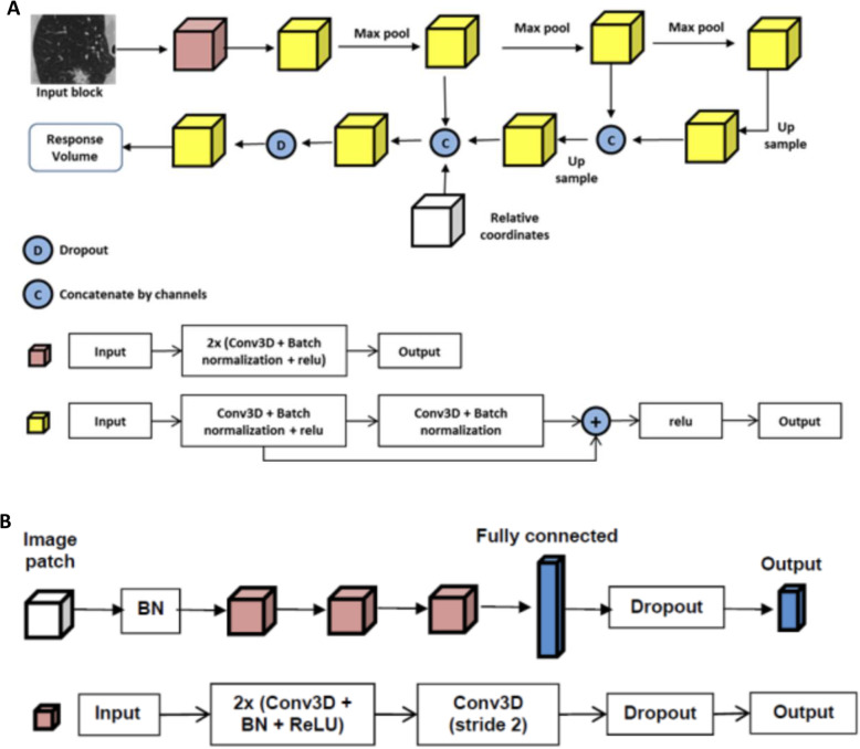
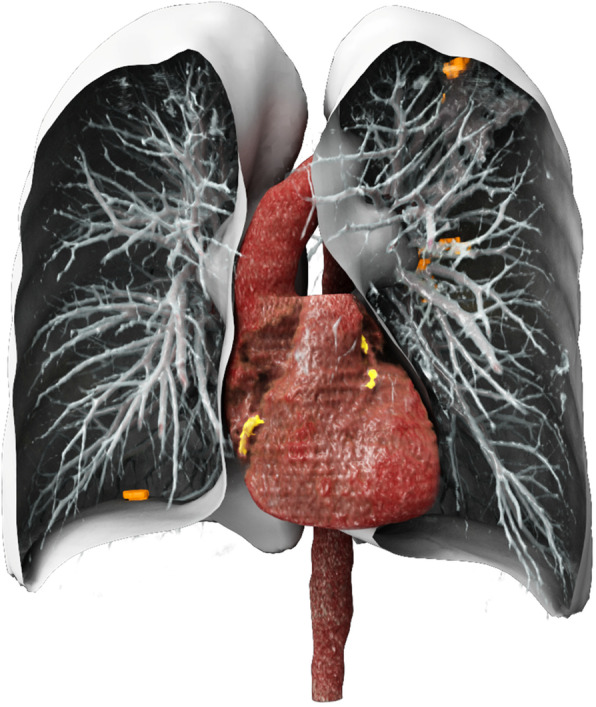
Artificial intelligence (AI) in diagnostic radiology is undergoing rapid development. Its potential utility to improve diagnostic performance for cardiopulmonary events is widely recognized, but the accuracy and precision have yet to be demonstrated in the context of current screening modalities. Here, we present findings on the performance of an AI convolutional neural network (CNN) prototype (AI-RAD Companion, Siemens Healthineers) that automatically detects pulmonary nodules and quantifies coronary artery calcium volume (CACV) on low-dose chest CT (LDCT), and compare results to expert radiologists. We also correlate AI findings with adverse cardiopulmonary outcomes in a retrospective cohort of 117 patients who underwent LDCT.
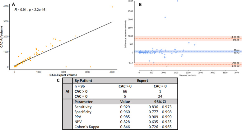
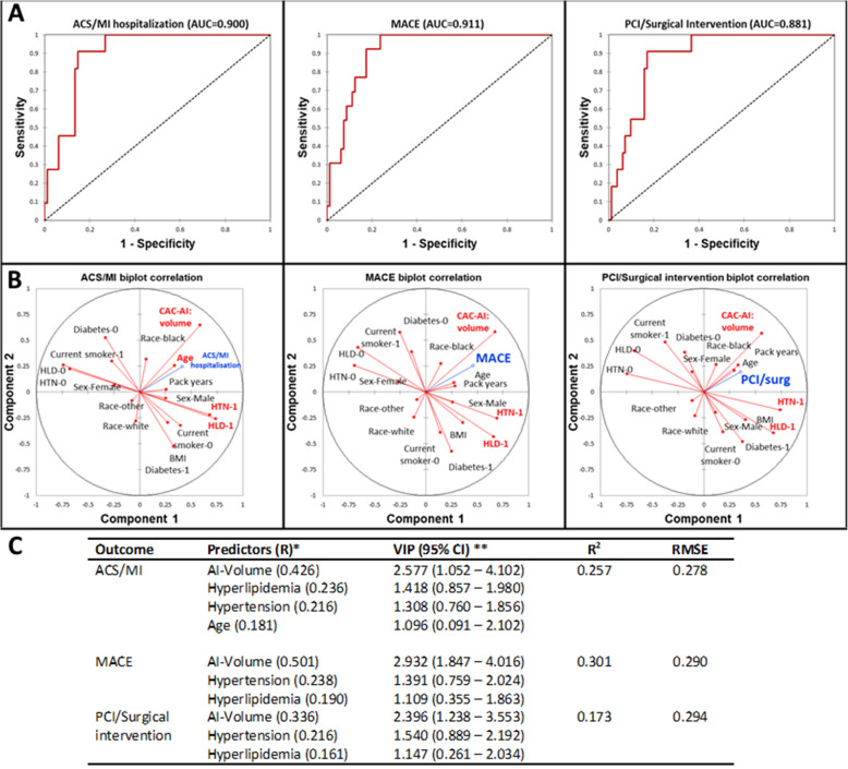
A total of 117 patients were enrolled in this study. Two CNNs were used to identify lung nodules and CACV on LDCT scans. All subjects were used for lung nodule analysis, and 96 subjects met the criteria for coronary artery calcium volume analysis. Interobserver concordance was measured using ICC and Cohen's kappa. Multivariate logistic regression and partial least squares regression were used for outcomes analysis.
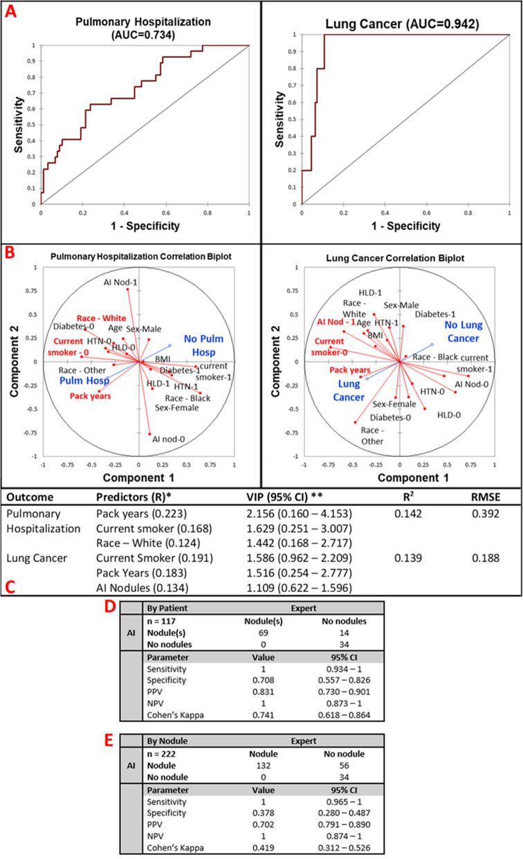
Agreement of the AI findings with experts was excellent (CACV ICC = 0.904, lung nodules Cohen's kappa = 0.846) with high sensitivity and specificity (CACV: sensitivity = .929, specificity = .960; lung nodules: sensitivity = 1, specificity = 0.708). The AI findings improved the prediction of major cardiopulmonary outcomes at 1-year follow-up including major adverse cardiac events and lung cancer (AUC = 0.911, AUC = 0.942).
We conclude the AI prototype rapidly and accurately identifies significant risk factors for cardiopulmonary disease on standard screening low-dose chest CT. This information can be used to improve diagnostic ability, facilitate intervention, improve morbidity and mortality, and decrease healthcare costs. There is also potential application in countries with limited numbers of cardiothoracic radiologists.
人工智能(AI)在诊断放射学领域发展迅速。人们广泛认识到其在改善心肺事件诊断性能方面的潜在效用,但在当前筛查模式下,其准确性和精密度尚未得到证实。在这里,我们展示了一种人工智能卷积神经网络(CNN)原型(AI-RAD Companion,西门子医疗)的性能研究结果,该原型可自动检测肺部结节并量化冠状动脉钙体积(CACV),并比较结果与专家放射科医生的结果。我们还在一个回顾性队列中对 117 名接受低剂量胸部 CT(LDCT)检查的患者的 AI 结果与不良心肺结局进行了相关性分析。
这项研究共纳入了 117 名患者。两个 CNN 用于在 LDCT 扫描中识别肺结节和 CACV。所有患者均用于肺结节分析,96 名患者符合冠状动脉钙体积分析标准。使用 ICC 和 Cohen's kappa 测量观察者间的一致性。使用多变量逻辑回归和偏最小二乘回归进行结果分析。
AI 结果与专家的一致性非常好(CACV 的 ICC=0.904,肺结节的 Cohen's kappa=0.846),具有较高的敏感性和特异性(CACV:敏感性=0.929,特异性=0.960;肺结节:敏感性=1,特异性=0.708)。AI 结果改善了 1 年随访期间主要心肺结局的预测,包括主要不良心脏事件和肺癌(AUC=0.911,AUC=0.942)。
我们的结论是,该 AI 原型可快速准确地识别标准筛查 LDCT 上与心肺疾病相关的重要危险因素。这些信息可用于提高诊断能力,促进干预,降低发病率和死亡率,并降低医疗保健成本。在心脏和胸部放射科医生数量有限的国家也有潜在的应用。