Shevchenko Victoria, Benn R Austin, Scholz Robert, Wei Wei, Pallavicini Carla, Klatzmann Ulysse, Alberti Francesco, Satterthwaite Theodore D, Wassermann Demian, Bazin Pierre-Louis, Margulies Daniel S
Cognitive Neuroanatomy Lab, INCC UMR 8002, CNRS, Université Paris Cité, Paris, France.
Wellcome Centre for Integrative Neuroimaging, Nuffield Department of Clinical Neurosciences, FMRIB Centre, University of Oxford, Oxford, UK.
Sci Rep. 2025 Jan 22;15(1):2849. doi: 10.1038/s41598-024-84152-2.
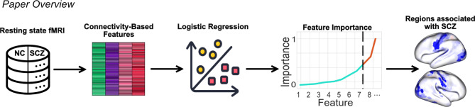
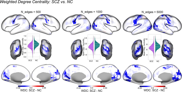
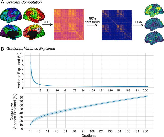
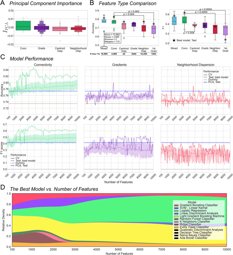
Functional connectivity holds promise as a biomarker of schizophrenia. Yet, the high dimensionality of predictive models trained on functional connectomes, combined with small sample sizes in clinical research, increases the risk of overfitting. Recently, low-dimensional representations of the connectome such as macroscale cortical gradients and gradient dispersion have been proposed, with studies noting consistent gradient and dispersion differences in psychiatric conditions. However, it is unknown which of these derived measures has the highest predictive capacity and how they compare to raw functional connectivity specifically in the case of schizophrenia. Our study evaluates which connectome features derived from resting state functional MRI - functional connectivity, gradients, or gradient dispersion - best identify schizophrenia. To this end, we leveraged data of 936 individuals from three large open-access datasets: COBRE, LA5c, and SRPBS-1600. We developed a pipeline which allows us to aggregate over a million different features and assess their predictive potential in a single, computationally efficient experiment. We selected top 1% of features with the largest permutation feature importance and trained 13 classifiers on them using 10-fold cross-validation. Our findings indicate that functional connectivity outperforms its low-dimensional derivatives such as cortical gradients and gradient dispersion in identifying schizophrenia (Mann-Whitney test conducted on test accuracy: connectivity vs. 1st gradient: U = 142, p < 0.003; connectivity vs. neighborhood dispersion: U = 141, p = 0.004). Additionally, we demonstrated that the edges which contribute the most to classification performance are the ones connecting primary sensory regions. Functional connectivity within the primary sensory regions showed the highest discrimination capabilities between subjects with schizophrenia and neurotypical controls. These findings along with the feature selection pipeline proposed here will facilitate future inquiries into the prediction of schizophrenia subtypes and transdiagnostic phenomena.
功能连接有望成为精神分裂症的一种生物标志物。然而,基于功能连接组训练的预测模型具有高维度性,再加上临床研究中的样本量较小,这增加了过拟合的风险。最近,有人提出了连接组的低维表示,如宏观皮层梯度和梯度离散度,研究指出在精神疾病中梯度和离散度存在一致的差异。然而,尚不清楚这些派生指标中哪一个具有最高的预测能力,以及它们与原始功能连接相比如何,特别是在精神分裂症的情况下。我们的研究评估了从静息态功能磁共振成像得出的哪些连接组特征——功能连接、梯度或梯度离散度——最能识别精神分裂症。为此,我们利用了来自三个大型开放获取数据集(COBRE、LA5c和SRPBS - 1600)的936名个体的数据。我们开发了一个流程,使我们能够汇总超过一百万个不同的特征,并在一个计算效率高的单一实验中评估它们的预测潜力。我们选择了排列特征重要性最大的前1%的特征,并使用10折交叉验证在这些特征上训练了13个分类器。我们的研究结果表明,在识别精神分裂症方面,功能连接优于其低维衍生物,如皮层梯度和梯度离散度(对测试准确率进行曼 - 惠特尼检验:连接性与一阶梯度:U = 142,p < 0.003;连接性与邻域离散度:U = 141,p = 0.004)。此外,我们证明了对分类性能贡献最大的边是连接主要感觉区域的边。主要感觉区域内的功能连接在精神分裂症患者和神经典型对照之间表现出最高的辨别能力。这些发现以及本文提出的特征选择流程将有助于未来对精神分裂症亚型和跨诊断现象预测的研究。