Teitz-Tennenbaum Seagal, Marinetti Kayla N, Lahiri Shayanki, Siddiqui Khadijah, Flory Celia, Tennenbaum Karinne, Hicks Helen G, Song Brian, Ganguly Anutosh, Osterholzer John J
Research Service and Pulmonary Section Medical Service, Veterans Affairs Ann Arbor Health System, Ann Arbor, Michigan, United States of America.
Division of Pulmonary and Critical Care Medicine, Department of Internal Medicine, University of Michigan, Ann Arbor, Michigan, United States of America.
PLoS One. 2025 Jan 24;20(1):e0313992. doi: 10.1371/journal.pone.0313992. eCollection 2025.
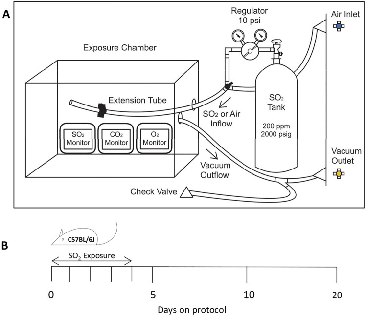
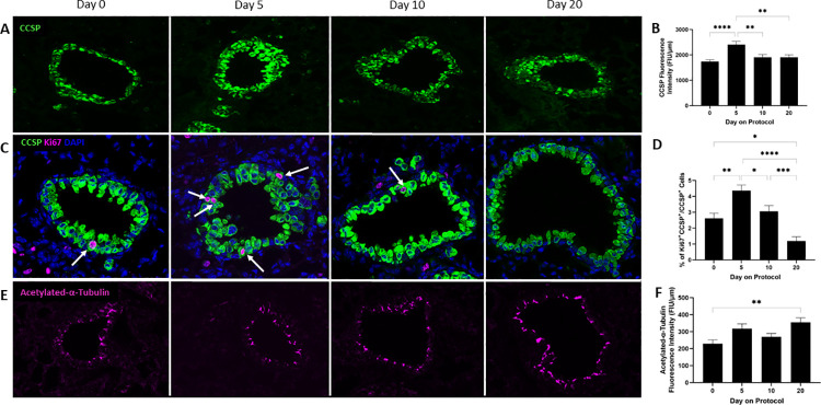
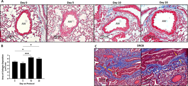
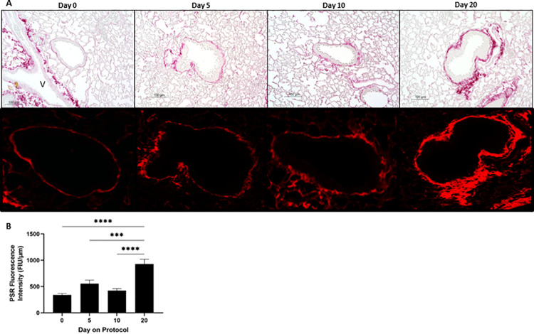
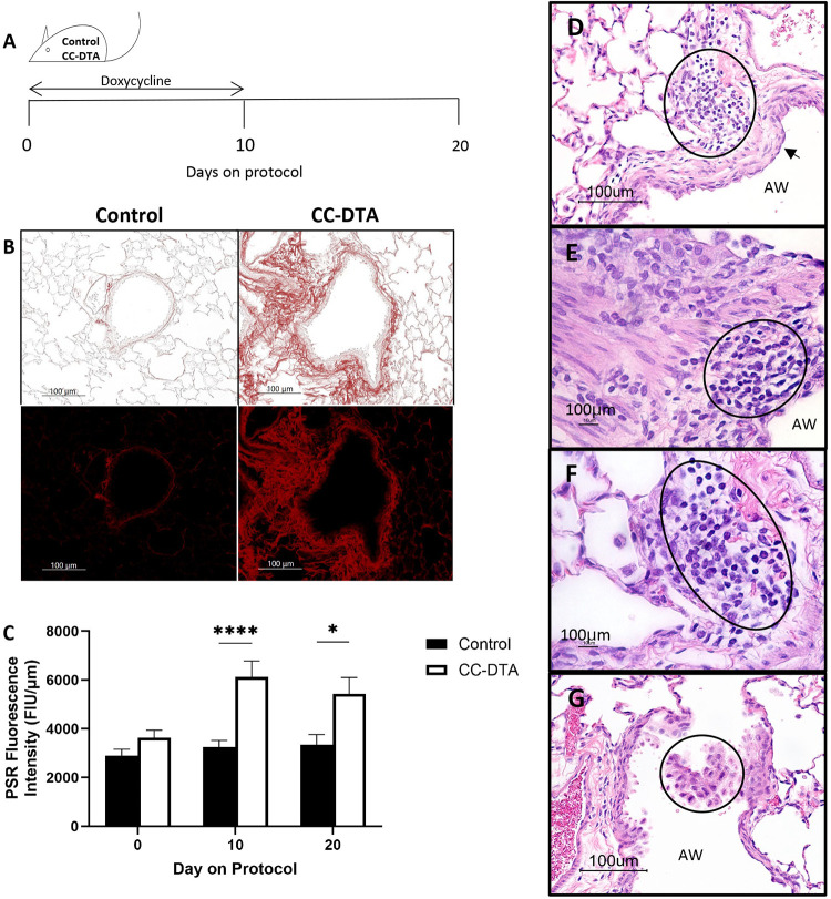
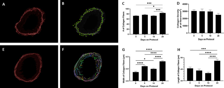
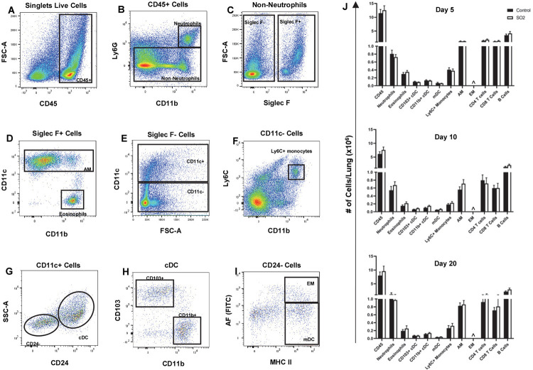
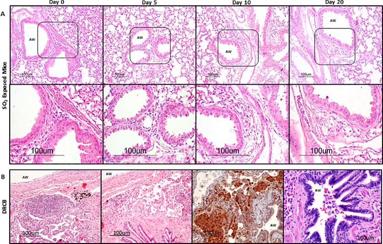
Deployment-related constrictive bronchiolitis (DRCB) has emerged as a health concern in military personnel returning from Southwest Asia. Exposure to smoke from a fire at the Al-Mishraq sulfur enrichment facility and/or burn pits was reported by a subset of Veterans diagnosed with this disorder. DRCB is characterized by thickening and fibrosis of small airways (SA) in the lung, but whether these are related to toxin inhalation remains uncertain. The aim of this study was to determine whether sulfur dioxide (SO2) exposure can induce histopathological features of DRCB. C57BL/6J mice were exposed to 50 ± 5 ppm SO2 for one hour/day for five consecutive days. Lungs from exposed and unexposed mice were evaluated on day 5, 10, and 20. Lung sections were stained using hematoxylin and eosin, Masson's trichrome, picrosirius red (PSR), and immunofluorescence for club cell secretory protein, acetylated-α-tubulin, and Ki67. Small airway wall thickness was determined by morphometric analysis and collagen content was quantified by measuring PSR fluorescence intensity. CurveAlign and CT-FIRE were used to enumerate collagen fibers and assess fibers' width and length, respectively. Leukocyte subpopulations were quantified by flow cytometry analysis. This protocol of SO2 exposure of mice: 1) Triggered club cell proliferation and differentiation; 2) Increased SA wall thickness by inducing subepithelial collagen deposition; and 3) Increased width, length, and number, but not density, of collagen fibers within the wall of SA. 4) Induced no peribronchiolar inflammation or respiratory bronchiolitis. Collectively, these findings implicate club cell proliferation and differentiation in the profibrotic response to SO2 and identify this SO2 exposure as a potentially effective though imperfect model for studying SA fibrosis in DRCB.
与部署相关的缩窄性细支气管炎(DRCB)已成为从西南亚返回的军事人员的一个健康问题。一部分被诊断患有这种疾病的退伍军人报告称,他们曾接触过米什拉克硫浓缩设施火灾产生的烟雾和/或燃烧坑。DRCB的特征是肺部小气道(SA)增厚和纤维化,但这些是否与毒素吸入有关仍不确定。本研究的目的是确定二氧化硫(SO₂)暴露是否会诱发DRCB的组织病理学特征。将C57BL/6J小鼠连续五天每天暴露于50±5 ppm的SO₂中1小时。在第5天、第10天和第20天对暴露和未暴露小鼠的肺部进行评估。肺切片用苏木精和伊红、Masson三色染色、苦味酸天狼星红(PSR)染色,并进行免疫荧光检测俱乐部细胞分泌蛋白、乙酰化α微管蛋白和Ki67。通过形态计量分析确定小气道壁厚度,并通过测量PSR荧光强度对胶原蛋白含量进行定量。分别使用CurveAlign和CT-FIRE来计数胶原纤维并评估纤维的宽度和长度。通过流式细胞术分析对白细胞亚群进行定量。这种SO₂暴露小鼠的方案:1)引发俱乐部细胞增殖和分化;2)通过诱导上皮下胶原沉积增加SA壁厚度;3)增加SA壁内胶原纤维的宽度、长度和数量,但不增加密度。4)未诱发细支气管周围炎症或呼吸性细支气管炎。总的来说,这些发现表明俱乐部细胞增殖和分化参与了对SO₂的促纤维化反应,并确定这种SO₂暴露是研究DRCB中SA纤维化的一个潜在有效但并不完美的模型。