Liu Yuming, Keikhosravi Adib, Pehlke Carolyn A, Bredfeldt Jeremy S, Dutson Matthew, Liu Haixiang, Mehta Guneet S, Claus Robert, Patel Akhil J, Conklin Matthew W, Inman David R, Provenzano Paolo P, Sifakis Eftychios, Patel Jignesh M, Eliceiri Kevin W
Laboratory for Optical and Computational Instrumentation, University of Wisconsin-Madison, Madison, WI, United States.
Department of Biomedical Engineering, University of Wisconsin-Madison, Madison, WI, United States.
Front Bioeng Biotechnol. 2020 Apr 21;8:198. doi: 10.3389/fbioe.2020.00198. eCollection 2020.
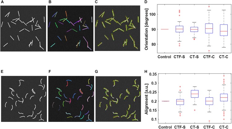
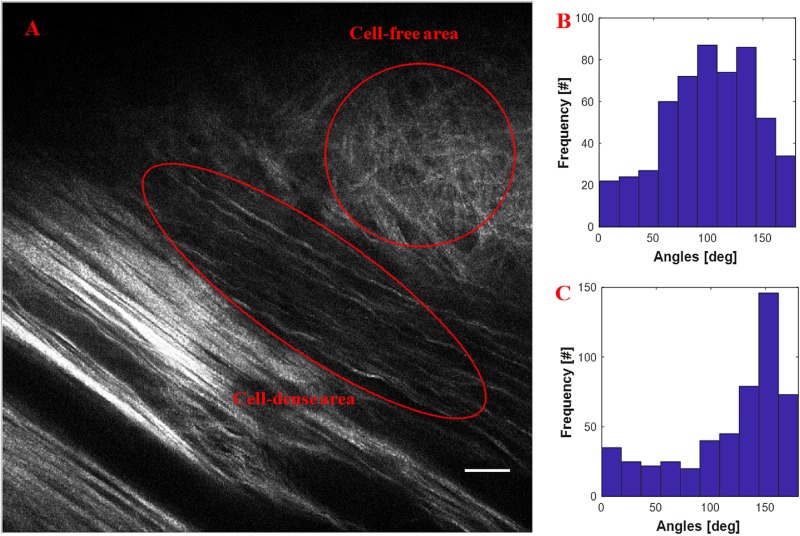
Quantification of fibrillar collagen organization has given new insight into the possible role of collagen topology in many diseases and has also identified candidate image-based bio-markers in breast cancer and pancreatic cancer. We have been developing collagen quantification tools based on the curvelet transform (CT) algorithm and have demonstrated this to be a powerful multiscale image representation method due to its unique features in collagen image denoising and fiber edge enhancement. In this paper, we present our CT-based collagen quantification software platform with a focus on new features and also giving a detailed description of curvelet-based fiber representation. These new features include C++-based code optimization for fast individual fiber tracking, Java-based synthetic fiber generator module for method validation, automatic tumor boundary generation for fiber relative quantification, parallel computing for large-scale batch mode processing, region-of-interest analysis for user-specified quantification, and pre- and post-processing modules for individual fiber visualization. We present a validation of the tracking of individual fibers and fiber orientations by using synthesized fibers generated by the synthetic fiber generator. In addition, we provide a comparison of the fiber orientation calculation on pancreatic tissue images between our tool and three other quantitative approaches. Lastly, we demonstrate the use of our software tool for the automatic tumor boundary creation and the relative alignment quantification of collagen fibers in human breast cancer pathology images, as well as the alignment quantification of mouse xenograft breast cancer images.
纤维状胶原蛋白组织的量化为胶原蛋白拓扑结构在多种疾病中的可能作用提供了新的见解,并且还在乳腺癌和胰腺癌中确定了基于图像的候选生物标志物。我们一直在基于曲波变换(CT)算法开发胶原蛋白量化工具,并已证明由于其在胶原蛋白图像去噪和纤维边缘增强方面的独特特性,它是一种强大的多尺度图像表示方法。在本文中,我们展示了基于CT的胶原蛋白量化软件平台,重点介绍新功能,并详细描述基于曲波的纤维表示。这些新功能包括用于快速单个纤维跟踪的基于C++的代码优化、用于方法验证的基于Java的合成纤维生成器模块、用于纤维相对量化的自动肿瘤边界生成、用于大规模批处理模式的并行计算、用于用户指定量化的感兴趣区域分析以及用于单个纤维可视化的预处理和后处理模块。我们通过使用合成纤维生成器生成的合成纤维展示了单个纤维及其取向跟踪的验证。此外,我们比较了我们的工具与其他三种定量方法在胰腺组织图像上的纤维取向计算。最后,我们展示了我们的软件工具在人类乳腺癌病理图像中自动创建肿瘤边界和胶原蛋白纤维相对排列量化,以及小鼠异种移植乳腺癌图像排列量化中的应用。