• 文献检索
  • 文档翻译
  • 深度研究
  • 学术资讯
  • Suppr Zotero 插件Zotero 插件
  • 邀请有礼
  • 套餐&价格
  • 历史记录
应用&插件
Suppr Zotero 插件Zotero 插件浏览器插件Mac 客户端Windows 客户端微信小程序
定价
高级版会员购买积分包购买API积分包
服务
文献检索文档翻译深度研究API 文档MCP 服务
关于我们
关于 Suppr公司介绍联系我们用户协议隐私条款
关注我们

Suppr 超能文献

核心技术专利:CN118964589B侵权必究
粤ICP备2023148730 号-1Suppr @ 2026

文献检索

告别复杂PubMed语法,用中文像聊天一样搜索,搜遍4000万医学文献。AI智能推荐,让科研检索更轻松。

立即免费搜索

文件翻译

保留排版,准确专业,支持PDF/Word/PPT等文件格式,支持 12+语言互译。

免费翻译文档

深度研究

AI帮你快速写综述,25分钟生成高质量综述,智能提取关键信息,辅助科研写作。

立即免费体验

通过深度学习实现面向临床的自动三维牙釉质分割

Clinically oriented automatic three-dimensional enamel segmentation via deep learning.

作者信息

Yu Wenting, Wang Xinwen, Yang Huifang

机构信息

Department of Orthodontics, School of Stomatology, Beijing Stomatological Hospital, Capital Medical University, Beijing, 100050, PR China.

Third Clinical Division, Peking University School and Hospital of Stomatology & National Center of Stomatology & National Clinical Research Center for Oral Diseases & National Engineering Research Center of Oral Biomaterials and Digital Medical Devices, Beijing, CN, China.

出版信息

BMC Oral Health. 2025 Jan 24;25(1):133. doi: 10.1186/s12903-024-05385-1.

DOI:10.1186/s12903-024-05385-1
PMID:39856656
原文链接:https://pmc.ncbi.nlm.nih.gov/articles/PMC11761753/
Abstract

BACKGROUND

Establishing accurate, reliable, and convenient methods for enamel segmentation and analysis is crucial for effectively planning endodontic, orthodontic, and restorative treatments, as well as exploring the evolutionary patterns of mammals. However, no mature, non-destructive method currently exists in clinical dentistry to quickly, accurately, and comprehensively assess the integrity and thickness of enamel chair-side. This study aims to develop a deep learning work, 2.5D Attention U-Net, trained on small sample datasets, for the automatical, efficient, and accurate segmentation of enamel across all teeth in clinical settings.

METHODS

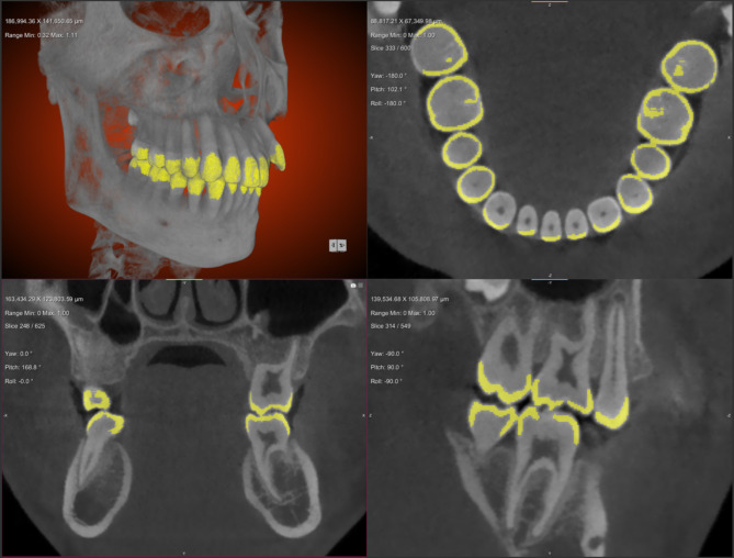
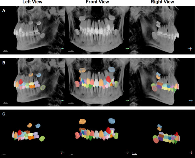
We propose a fully automated computer-aided enamel segmentation model based on an instance segmentation network, 2.5D Attention U-Net. After data annotation and augmentation, the model is trained using manually annotated segmented enamel data, and its performance is evaluated using the Dice similarity coefficient metrics. A satisfactory image segmentation model is applied to generate a 3D enamel model for each tooth and to calculate the thickness value of individual enclosed 3D enamel meshes using a normal ray-tracing directional method.

RESULTS

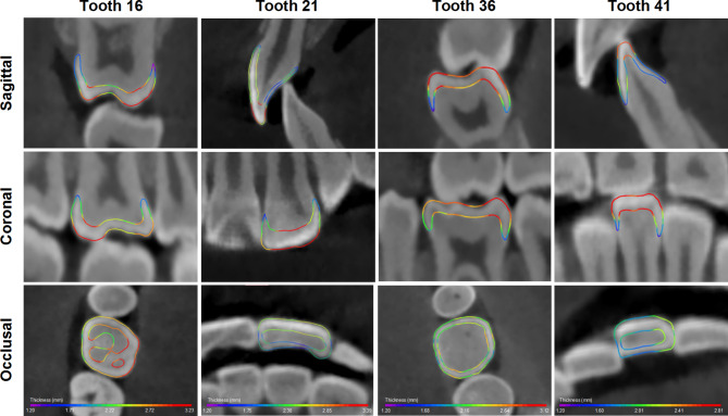
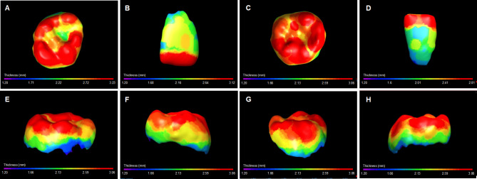
The model achieves the Dice score on the enamel segmentation task of 96.6%. This study provides an intuitive visualization of irregular enamel morphology and a quantitative analysis of three-dimensional enamel thickness variations. The results indicate that enamel is thickest at the incisal edges of anterior teeth and the cusps of posterior teeth, thinning towards the roots. For posterior teeth, the enamel is thinnest at the central fossae area, with mandibular molars having thicker enamel in the central fossae compared to maxillary molars. The average enamel thickness of maxillary incisors, canines, and premolars is greater than that of mandibular incisors, while the opposite is true for molars. Although there are individual variations in enamel thickness, the average enamel thickness graduallly increases from the incisors to the molars among all teeth within the same quadrant.

CONCLUSIONS

This study introduces an automatic, efficient, and accurate 2.5D Attention U-Net system to enhance precise and efficient chair-side diagnosis and treatment of enamel-related diseases in clinical settings, marking a significant advancement in automated diagnostics for enamel-related conditions.

摘要

背景

建立准确、可靠且便捷的牙釉质分割与分析方法,对于有效规划牙髓病、正畸和修复治疗,以及探索哺乳动物的进化模式至关重要。然而,目前临床牙科中尚无成熟的非破坏性方法能够在椅旁快速、准确且全面地评估牙釉质的完整性和厚度。本研究旨在开发一种深度学习模型——2.5D注意力U-Net,在小样本数据集上进行训练,以在临床环境中自动、高效且准确地分割所有牙齿的牙釉质。

方法

我们提出了一种基于实例分割网络2.5D注意力U-Net的全自动计算机辅助牙釉质分割模型。在数据标注和增强后,使用手动标注的分割牙釉质数据对模型进行训练,并使用Dice相似系数指标评估其性能。应用一个令人满意的图像分割模型为每颗牙齿生成三维牙釉质模型,并使用法线光线追踪定向方法计算各个封闭三维牙釉质网格的厚度值。

结果

该模型在牙釉质分割任务上的Dice分数达到96.6%。本研究提供了不规则牙釉质形态的直观可视化以及三维牙釉质厚度变化的定量分析。结果表明,牙釉质在前牙切缘和后牙牙尖处最厚,向牙根方向逐渐变薄。对于后牙,牙釉质在中央窝区域最薄,下颌磨牙中央窝处牙釉质比上颌磨牙更厚。上颌切牙、尖牙和前磨牙的平均牙釉质厚度大于下颌切牙,而磨牙则相反。尽管牙釉质厚度存在个体差异,但同一象限内所有牙齿的平均牙釉质厚度从切牙到磨牙逐渐增加。

结论

本研究引入了一种自动、高效且准确地2.5D注意力U-Net系统,以加强临床环境中牙釉质相关疾病的精确和高效椅旁诊断与治疗,标志着牙釉质相关病症自动诊断方面的重大进展。

https://cdn.ncbi.nlm.nih.gov/pmc/blobs/2cc2/11761753/46c267619a95/12903_2024_5385_Fig6_HTML.jpg
https://cdn.ncbi.nlm.nih.gov/pmc/blobs/2cc2/11761753/4d1b24c21ffe/12903_2024_5385_Fig1_HTML.jpg
https://cdn.ncbi.nlm.nih.gov/pmc/blobs/2cc2/11761753/2136f6fa31d2/12903_2024_5385_Fig2_HTML.jpg
https://cdn.ncbi.nlm.nih.gov/pmc/blobs/2cc2/11761753/7e37a3cdd84d/12903_2024_5385_Fig3_HTML.jpg
https://cdn.ncbi.nlm.nih.gov/pmc/blobs/2cc2/11761753/2eab80156145/12903_2024_5385_Fig4_HTML.jpg
https://cdn.ncbi.nlm.nih.gov/pmc/blobs/2cc2/11761753/895235dfccf5/12903_2024_5385_Fig5_HTML.jpg
https://cdn.ncbi.nlm.nih.gov/pmc/blobs/2cc2/11761753/46c267619a95/12903_2024_5385_Fig6_HTML.jpg

相似文献

1
Clinically oriented automatic three-dimensional enamel segmentation via deep learning.通过深度学习实现面向临床的自动三维牙釉质分割
BMC Oral Health. 2025 Jan 24;25(1):133. doi: 10.1186/s12903-024-05385-1.
2
Fully automated method for three-dimensional segmentation and fine classification of mixed dentition in cone-beam computed tomography using deep learning.基于深度学习的锥形束计算机断层扫描中混合牙列的全自动三维分割和精细分类方法。
J Dent. 2024 Dec;151:105398. doi: 10.1016/j.jdent.2024.105398. Epub 2024 Oct 22.
3
Accurate object localization facilitates automatic esophagus segmentation in deep learning.准确的目标定位有助于深度学习中的自动食管分割。
Radiat Oncol. 2024 May 12;19(1):55. doi: 10.1186/s13014-024-02448-z.
4
Automatic prostate segmentation using deep learning on clinically diverse 3D transrectal ultrasound images.基于临床多样的三维经直肠超声图像,利用深度学习进行前列腺自动分割。
Med Phys. 2020 Jun;47(6):2413-2426. doi: 10.1002/mp.14134. Epub 2020 Apr 8.
5
Deep learning for automated segmentation of the temporomandibular joint.用于颞下颌关节自动分割的深度学习
J Dent. 2023 May;132:104475. doi: 10.1016/j.jdent.2023.104475. Epub 2023 Mar 2.
6
A novel deep learning system for multi-class tooth segmentation and classification on cone beam computed tomography. A validation study.一种基于锥形束 CT 的新型深度学习多类牙分割与分类系统:验证研究。
J Dent. 2021 Dec;115:103865. doi: 10.1016/j.jdent.2021.103865. Epub 2021 Oct 26.
7
Brain tumor segmentation and detection in MRI using convolutional neural networks and VGG16.使用卷积神经网络和VGG16在磁共振成像(MRI)中进行脑肿瘤分割与检测
Cancer Biomark. 2025 Mar;42(3):18758592241311184. doi: 10.1177/18758592241311184. Epub 2025 Apr 4.
8
Technical Note: Guidelines for the digital computation of 2D and 3D enamel thickness in hominoid teeth.技术说明:人科牙齿二维和三维釉质厚度数字计算指南。
Am J Phys Anthropol. 2014 Feb;153(2):305-13. doi: 10.1002/ajpa.22421. Epub 2013 Nov 18.
9
Enamel thickness in primary teeth.乳牙的牙釉质厚度
J Clin Pediatr Dent. 2012 Winter;37(2):177-81. doi: 10.17796/jcpd.37.2.d6837416076l3334.
10
A robust and automatic CT-3D ultrasound registration method based on segmentation, context, and edge hybrid metric.基于分割、上下文和边缘混合度量的稳健自动 CT-3D 超声配准方法。
Med Phys. 2023 Oct;50(10):6243-6258. doi: 10.1002/mp.16396. Epub 2023 Apr 6.

本文引用的文献

1
In Vitro Analysis of Enamel Patterns Across Three Species Using Stereomicroscopy.使用体视显微镜对三个物种的牙釉质形态进行体外分析。
Cureus. 2024 May 1;16(5):e59488. doi: 10.7759/cureus.59488. eCollection 2024 May.
2
Fully automatic AI segmentation of oral surgery-related tissues based on cone beam computed tomography images.基于锥形束计算机断层扫描图像的口腔外科相关组织全自动 AI 分割。
Int J Oral Sci. 2024 May 8;16(1):34. doi: 10.1038/s41368-024-00294-z.
3
A progressive framework for tooth and substructure segmentation from cone-beam CT images.
基于锥形束 CT 图像的牙齿及子结构渐进式分割框架。
Comput Biol Med. 2024 Feb;169:107839. doi: 10.1016/j.compbiomed.2023.107839. Epub 2023 Dec 13.
4
Age and sex related change in tooth enamel thickness of maxillary incisors measured by cone beam computed tomography.应用锥形束 CT 测量上颌中切牙牙釉质厚度的年龄和性别相关变化。
BMC Oral Health. 2023 Dec 6;23(1):971. doi: 10.1186/s12903-023-03639-y.
5
Mammalian tooth enamel functional sophistication demonstrated by combined nanotribology and synchrotron radiation FTIR analyses.通过纳米摩擦学与同步辐射傅里叶变换红外光谱分析相结合所展示的哺乳动物牙釉质功能复杂性。
iScience. 2022 Dec 27;26(1):105679. doi: 10.1016/j.isci.2022.105679. eCollection 2023 Jan 20.
6
Influence of water and protein content on the creep behavior in dental enamel.水和蛋白质含量对牙釉质蠕变行为的影响。
Acta Biomater. 2023 Mar 1;158:393-411. doi: 10.1016/j.actbio.2023.01.018. Epub 2023 Jan 11.
7
A fully automatic AI system for tooth and alveolar bone segmentation from cone-beam CT images.基于锥形束 CT 图像的全自动 AI 牙齿和牙槽骨分割系统。
Nat Commun. 2022 Apr 19;13(1):2096. doi: 10.1038/s41467-022-29637-2.
8
Proximal enamel thickness of the permanent teeth: A systematic review and meta-analysis.恒牙近釉质厚度的系统评价和荟萃分析。
Am J Orthod Dentofacial Orthop. 2021 Dec;160(6):793-804.e3. doi: 10.1016/j.ajodo.2021.05.007. Epub 2021 Aug 19.
9
Transillumination and optical coherence tomography for the detection and diagnosis of enamel caries.透照和光相干断层扫描在釉质龋的检测和诊断中的应用。
Cochrane Database Syst Rev. 2021 Jan 27;1(1):CD013855. doi: 10.1002/14651858.CD013855.
10
Enamel thickness of maxillary canines evaluated with microcomputed tomography scans.上颌尖牙釉质厚度的微计算机断层扫描评估。
Am J Orthod Dentofacial Orthop. 2020 Sep;158(3):391-399. doi: 10.1016/j.ajodo.2019.09.013. Epub 2020 Jul 9.