Wang Qiaolin, Wu Yutong, Ouyang Lianlian, Min Xiaoli, Zheng Meiling, Gao Lingyu, Chen Xiaoyun, Hu Zhi, Yang Shuang, Jiang Wenjuan, Jia Sujie, Lu Qianjin, Zhao Ming
Hospital for Skin Diseases, Institute of Dermatology, Chinese Academy of Medical Sciences and Peking Union Medical College, Nanjing, 210042, China.
Key Laboratory of Basic and Translational Research on Immune-Mediated Skin Diseases, Chinese Academy of Medical Sciences, Nanjing, 210042, China.
J Transl Med. 2025 Jan 27;23(1):118. doi: 10.1186/s12967-025-06147-5.
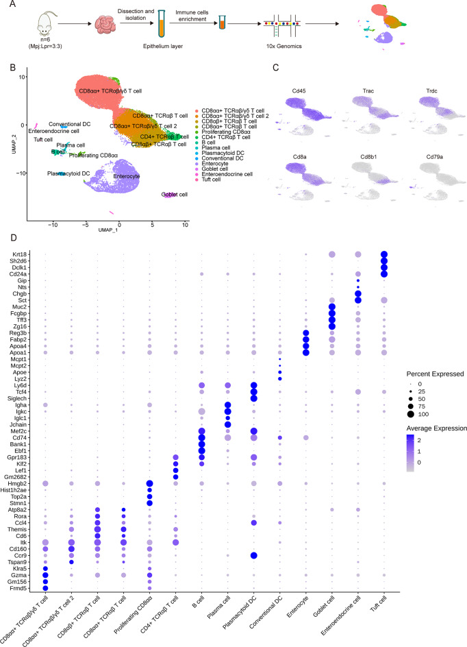
The small intestine harbors a rich array of intestinal intraepithelial lymphocytes (IELs) that interact with structural cells to collectively sustain gut immune homeostasis. Dysregulation of gut immune homeostasis was implicated in the pathogenesis of multiple autoimmune diseases, however, whether this homeostasis is disrupted in a lupus autoimmune background remains unclear.
We performed single-cell RNA sequencing (scRNA-seq) analyses to elucidate immune and structural milieu in the intestinal epithelium of MRL/Lpr lupus mice (Lpr mice) and MRL/Mpj control mice (Mpj mice). Comprehensive analyses including unsupervised clustering, trajectories, and cellular communication were performed. The primary findings from scRNA-seq were further validated by quantitative polymerase chain reaction (qPCR), flow cytometry, and in vivo experiments including selenium supplementation.
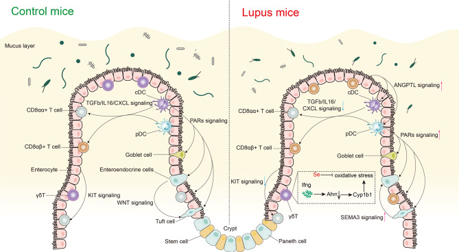
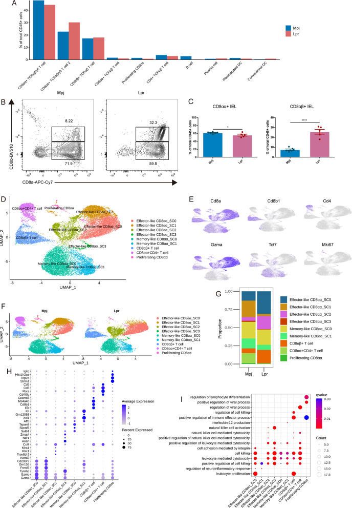
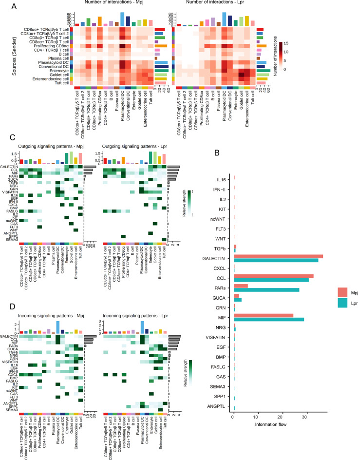
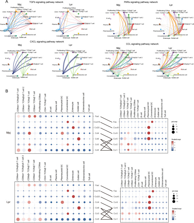
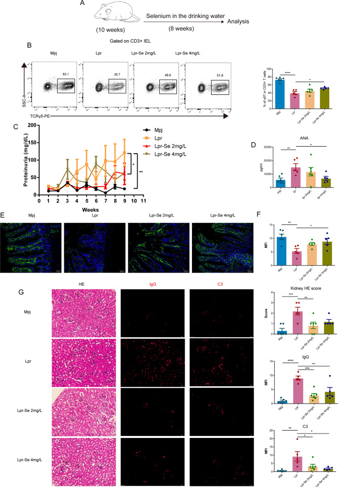
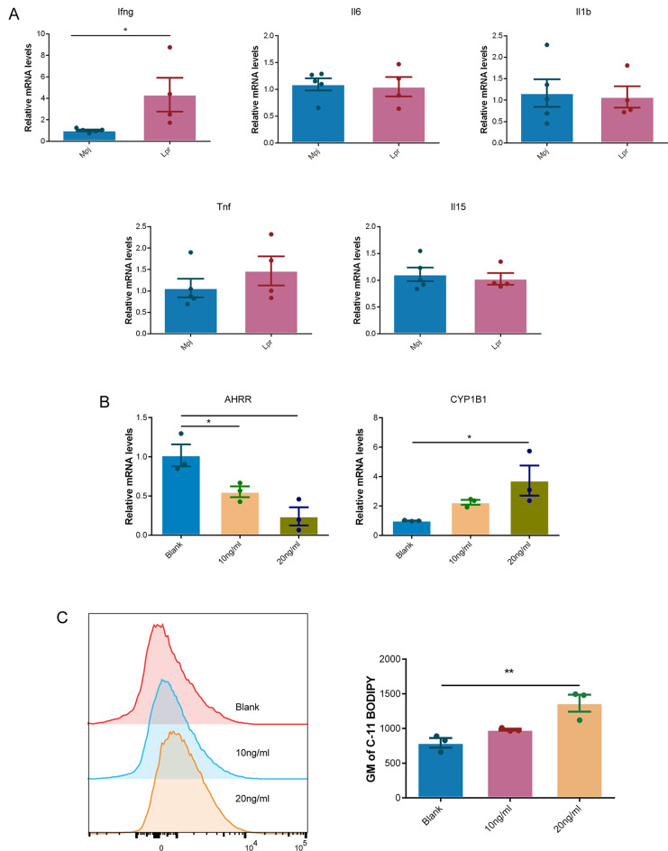
We observed a significant reduction in CD8αα + IELs, accompanied by a marked increase in CD8αβ + IELs in Lpr mice. Additionally, subsets of CD8 + IELs exhibiting significantly enhanced effector functions were found to be markedly enriched in Lpr mice. Intercellular communication patterns within intestinal epithelial immune and structural cells were found to be specifically altered in Lpr mice. Moreover, scRNA-seq revealed significantly decreased intestinal TCRγδ T cells (γδT) associated with reduced aryl-hydrocarbon receptor repressor (AHRR) expression and subsequent oxidative stress and ferroptosis in Lpr mice. Antioxidant selenium effectively reversed the loss of γδT in Lpr mice, improved the gut barrier, and alleviated lupus symptoms.
Our high-resolution single-cell atlas enhances the understanding of the immune and structural milieu of intestinal epithelium in lupus and provides new insights into lupus pathogenesis mediated by intestinal immune dysregulation.
小肠中含有大量肠道上皮内淋巴细胞(IELs),它们与结构细胞相互作用,共同维持肠道免疫稳态。肠道免疫稳态失调与多种自身免疫性疾病的发病机制有关,然而,在狼疮自身免疫背景下这种稳态是否被破坏仍不清楚。
我们进行了单细胞RNA测序(scRNA-seq)分析,以阐明MRL/Lpr狼疮小鼠(Lpr小鼠)和MRL/Mpj对照小鼠(Mpj小鼠)肠道上皮中的免疫和结构环境。进行了包括无监督聚类、轨迹分析和细胞通讯在内的综合分析。scRNA-seq的主要发现通过定量聚合酶链反应(qPCR)、流式细胞术以及包括补充硒在内的体内实验进一步验证。
我们观察到Lpr小鼠中CD8αα+ IELs显著减少,同时CD8αβ+ IELs显著增加。此外,发现Lpr小鼠中表现出效应功能显著增强的CD8+ IELs亚群明显富集。发现Lpr小鼠肠道上皮免疫和结构细胞内的细胞通讯模式发生了特异性改变。此外,scRNA-seq显示Lpr小鼠中肠道TCRγδ T细胞(γδT)显著减少,这与芳烃受体阻遏物(AHRR)表达降低以及随后的氧化应激和铁死亡有关。抗氧化剂硒有效地逆转了Lpr小鼠中γδT的损失,改善了肠道屏障,并减轻了狼疮症状。
我们的高分辨率单细胞图谱加深了对狼疮中肠道上皮免疫和结构环境的理解,并为肠道免疫失调介导的狼疮发病机制提供了新的见解。