León Oswaldo Cruz, Anzaldúa Arce Santiago René, Cornejo Cortes Miguel Angel, Cerbón Marco, Castañeda Francisco Ernesto Martínez, Arvizu Raúl Ulloa, Trujillo Ortega María Elena
Morphology Department, Facultad de Medicina Veterinaria y Zootecnia, National Autonomous University of Mexico, Mexico City, Mexico.
Biological Sciences Department, Facultad de Estudios Superiores Cuautitlán, National Autonomous University of Mexico, Cuautitlán Izcalli, State of Mexico, Mexico.
Anat Histol Embryol. 2025 Mar;54(2):e70017. doi: 10.1111/ahe.70017.
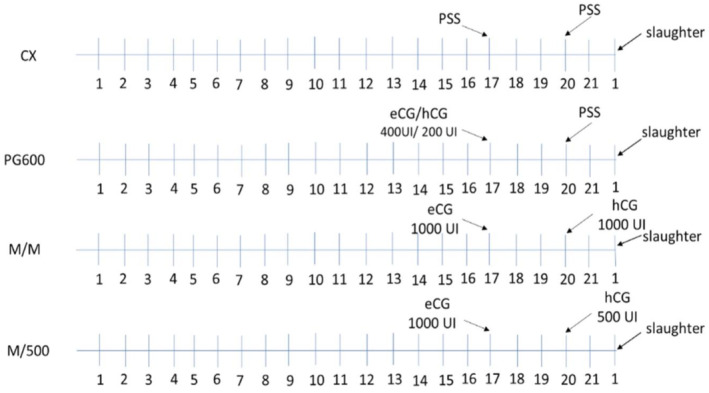
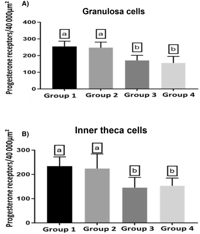
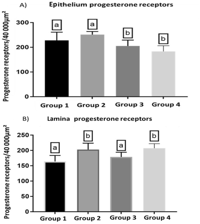
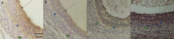
We studied sows (Landrace × Yorkshire line, DanBred Hybrid) to evaluate the possible changes in progesterone receptor (PR) expression in the uterus and ovary caused by different non-hypophyseal gonadotropins treatments: equine chorionic gonadotropin (eCG) and human chorionic gonadotropin (hCG). Varying concentrations of eCG and hCG were evaluated (Groups 1, 2, 3, 4). PR expression was determined by immunohistochemistry, and labelling intensity was determined by the HScore method. In the ovary, PR expression in the granulosa cells of follicles did not differ significantly between Groups 1 and 2 (p < 0.05) but differed significantly from that in Groups 3 and 4 (p < 0.05), which in turn did not differ from each other. This PR expression pattern was similar across groups in the internal and external theca cells. Conversely, in the uterus, PR expression in the lining epithelium was lower in Group 4 than that in Group 1 (p < 0.05). Increased expression was observed in the endometrial lamina propria in all groups 2 and 4 compared to that in the control group (p < 0.05). Decreased expression was observed in the glandular epithelium and myometrium in Group 4 compared to that in Group 1 (p < 0.05). In the ovary, PR expression in the granulosa and outer and inner theca of the follicles was not significantly different (p < 0.05) between Groups 1 and 2 or Groups 3 and 4; however, the expression in these pairs of groups differed from each other. Thus, changes in PR expression may depend on the concentrations and proportions of exogenous hormones used in the treatments, indicating an alteration in the reproductive process.
我们研究了母猪(长白猪×约克夏品系,DanBred杂种猪),以评估不同非垂体促性腺激素处理(马绒毛膜促性腺激素(eCG)和人绒毛膜促性腺激素(hCG))对子宫和卵巢中孕酮受体(PR)表达可能产生的变化。评估了不同浓度的eCG和hCG(第1、2、3、4组)。通过免疫组织化学测定PR表达,并通过HScore方法测定标记强度。在卵巢中,第1组和第2组卵泡颗粒细胞中的PR表达无显著差异(p<0.05),但与第3组和第4组有显著差异(p<0.05),而第3组和第4组之间无差异。这种PR表达模式在内外膜细胞的各组中相似。相反,在子宫中,第4组内膜上皮中的PR表达低于第1组(p<0.05)。与对照组相比,第2组和第4组所有组的子宫内膜固有层中均观察到表达增加(p<0.05)。与第1组相比,第4组腺上皮和子宫肌层中的表达降低(p<0.05)。在卵巢中,第1组和第2组或第3组和第4组之间卵泡颗粒细胞以及卵泡外层和内层膜中的PR表达无显著差异(p<0.05);然而,这两组之间的表达彼此不同。因此,PR表达的变化可能取决于处理中使用的外源激素的浓度和比例,表明生殖过程发生了改变。