• 文献检索
  • 文档翻译
  • 深度研究
  • 学术资讯
  • Suppr Zotero 插件Zotero 插件
  • 邀请有礼
  • 套餐&价格
  • 历史记录
应用&插件
Suppr Zotero 插件Zotero 插件浏览器插件Mac 客户端Windows 客户端微信小程序
定价
高级版会员购买积分包购买API积分包
服务
文献检索文档翻译深度研究API 文档MCP 服务
关于我们
关于 Suppr公司介绍联系我们用户协议隐私条款
关注我们

Suppr 超能文献

核心技术专利:CN118964589B侵权必究
粤ICP备2023148730 号-1Suppr @ 2026

文献检索

告别复杂PubMed语法,用中文像聊天一样搜索,搜遍4000万医学文献。AI智能推荐,让科研检索更轻松。

立即免费搜索

文件翻译

保留排版,准确专业,支持PDF/Word/PPT等文件格式,支持 12+语言互译。

免费翻译文档

深度研究

AI帮你快速写综述,25分钟生成高质量综述,智能提取关键信息,辅助科研写作。

立即免费体验

皮肤良性皮肤、软组织及附属器肿瘤的临床皮肤镜和组织病理学评估:一项来自三级医疗医院的横断面研究

Clinico-dermoscopic and Histopathological Evaluation of Benign Skin, Soft Tissue and Appendageal Tumours of Skin: A Cross-sectional Study from a Tertiary Care Hospital.

作者信息

Reyaz Saika, Rather Shagufta, Bilal Sheikh, Trumboo Taiba, Hussain Mateen

机构信息

From the Department of Dermatology, Government Medical College, Srinagar and Jammu, India.

Department of Pathology, Government Medical College, Srinagar, University of Kashmir, Jammu and Kashmir, India.

出版信息

Indian J Dermatol. 2025 Jan-Feb;70(1):50. doi: 10.4103/ijd.ijd_461_24. Epub 2024 Dec 30.

DOI:10.4103/ijd.ijd_461_24
PMID:39896304
原文链接:https://pmc.ncbi.nlm.nih.gov/articles/PMC11784977/
Abstract

BACKGROUND

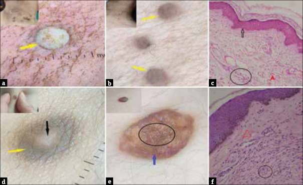
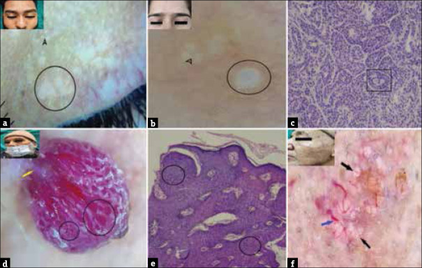
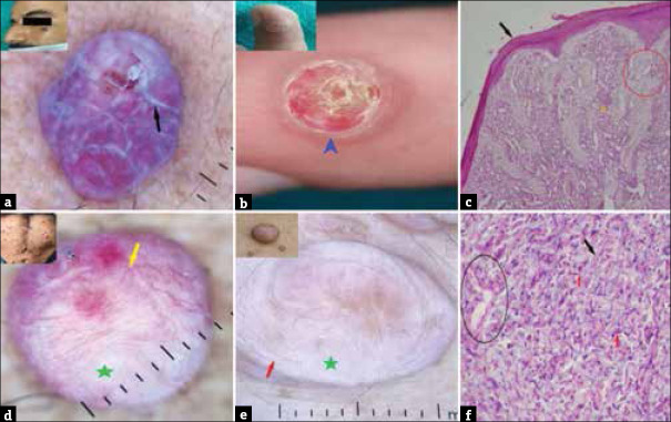
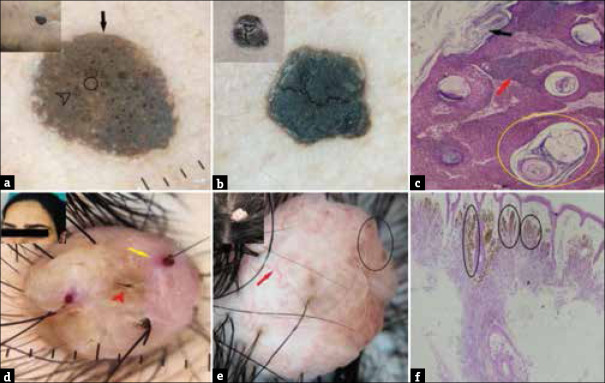
Benign skin, soft tissue and appendageal tumours of skin are one of the most frequently encountered skin disorders. An organised systematic approach along with dermoscopic and histopathological examination can aid in the diagnosis of these diverse disorders.

OBJECTIVE

To evaluate clinico-dermoscopic and histopathological findings of benign skin, soft tissue and appendageal tumours of skin in patients attending a tertiary care hospital.

METHODS

Cross-sectional hospital-based study where patients of all age groups irrespective of gender suspected of having benign skin, soft tissue and appendageal tumours were enrolled. Clinical, dermoscopic and histopathological findings were recorded and the agreements between them were evaluated using the Cohen's Kappa coefficient.

RESULTS

The study included a total of 415 patients with a mean age of 37.9 ± 15.59 years and a male to female ratio of 1:1.3. The mean duration of the disease was 4.3 ± 2.14 years. Soft tissue tumours were the commonest (60%), followed by benign skin tumours (24.3%) and benign appendageal tumours (17.1%). A good agreement between dermoscopic and clinical diagnosis was found (Cohen's Kappa = 0.879) and between dermoscopic and histopathological diagnosis was also found (Cohen's Kappa = 0.789).

CONCLUSION

This study infers that benign tumours of the skin include a heterogeneous group of skin disorders affecting a heterogeneous population. Dermoscopy improved the diagnostic accuracy of this large group of skin disorders and reduced the number of unnecessary excisions; however, histopathology remains the benchmark diagnostic tool to differentiate these tumours from other benign tumours and their malignant counterparts.

摘要

背景

皮肤良性肿瘤、软组织肿瘤及皮肤附属器肿瘤是最常见的皮肤疾病之一。采用系统有序的方法并结合皮肤镜检查和组织病理学检查有助于诊断这些多样的疾病。

目的

评估在一家三级医院就诊的患者中皮肤良性肿瘤、软组织肿瘤及皮肤附属器肿瘤的临床皮肤镜及组织病理学表现。

方法

基于医院的横断面研究,纳入所有年龄组、性别不限且疑似患有皮肤良性肿瘤、软组织肿瘤及皮肤附属器肿瘤的患者。记录临床、皮肤镜及组织病理学表现,并使用科恩kappa系数评估它们之间的一致性。

结果

该研究共纳入415例患者,平均年龄为37.9±15.59岁,男女比例为1:1.3。疾病平均病程为4.3±2.14年。软组织肿瘤最为常见(60%),其次是皮肤良性肿瘤(24.3%)和皮肤附属器良性肿瘤(17.1%)。皮肤镜与临床诊断之间具有良好的一致性(科恩kappa系数=0.879),皮肤镜与组织病理学诊断之间也具有良好的一致性(科恩kappa系数=0.789)。

结论

本研究推断皮肤良性肿瘤包括一组异质性的皮肤疾病,影响着异质性人群。皮肤镜提高了这一大组皮肤疾病的诊断准确性,减少了不必要的切除手术数量;然而,组织病理学仍然是区分这些肿瘤与其他良性肿瘤及其恶性对应物的基准诊断工具。

https://cdn.ncbi.nlm.nih.gov/pmc/blobs/ed80/11784977/a3c34aaff882/IJD-70-50b-g005.jpg
https://cdn.ncbi.nlm.nih.gov/pmc/blobs/ed80/11784977/8dc203ab4e45/IJD-70-50b-g001.jpg
https://cdn.ncbi.nlm.nih.gov/pmc/blobs/ed80/11784977/8685046084b0/IJD-70-50b-g002.jpg
https://cdn.ncbi.nlm.nih.gov/pmc/blobs/ed80/11784977/2070e7444ca1/IJD-70-50b-g003.jpg
https://cdn.ncbi.nlm.nih.gov/pmc/blobs/ed80/11784977/e5555bf783df/IJD-70-50b-g004.jpg
https://cdn.ncbi.nlm.nih.gov/pmc/blobs/ed80/11784977/a3c34aaff882/IJD-70-50b-g005.jpg

相似文献

1
Clinico-dermoscopic and Histopathological Evaluation of Benign Skin, Soft Tissue and Appendageal Tumours of Skin: A Cross-sectional Study from a Tertiary Care Hospital.皮肤良性皮肤、软组织及附属器肿瘤的临床皮肤镜和组织病理学评估:一项来自三级医疗医院的横断面研究
Indian J Dermatol. 2025 Jan-Feb;70(1):50. doi: 10.4103/ijd.ijd_461_24. Epub 2024 Dec 30.
2
Clinico-pathological Study of Skin Appendageal Tumours: An Experience at a Tertiary Care Hospital.皮肤附属器肿瘤的临床病理研究:一家三级护理医院的经验
Indian J Surg Oncol. 2024 Dec;15(4):625-632. doi: 10.1007/s13193-024-01963-5. Epub 2024 May 24.
3
Implications of Dermoscopy and Histopathological Correlation in Discoid Lupus Erythematosus in Skin of Color.皮肤镜检查与组织病理学相关性在有色人种盘状红斑狼疮中的意义
Indian J Dermatol. 2022 Jan-Feb;67(1):5-11. doi: 10.4103/ijd.ijd_591_21.
4
Correlation with digital dermoscopic images can help dermatopathologists to diagnose equivocal skin tumours.与数字皮肤镜图像的相关性有助于皮肤病理学家诊断不明确的皮肤肿瘤。
Br J Dermatol. 2006 Sep;155(3):546-51. doi: 10.1111/j.1365-2133.2006.07342.x.
5
An Observational Study of Dermoscopic and Histopathological Correlation in Spongiotic Disorders - A Hospital Based Cross Sectional Study.苔藓样变疾病的皮肤镜与组织病理学相关性观察研究——一项基于医院的横断面研究
Indian J Dermatol. 2023 Nov-Dec;68(6):634-641. doi: 10.4103/ijd.ijd_469_23. Epub 2024 Jan 9.
6
The use of dermoscopy in distinguishing the histopathological subtypes of basal cell carcinoma: A retrospective, morphological study.皮肤镜在鉴别基底细胞癌组织病理学亚型中的应用:一项回顾性形态学研究。
Indian J Dermatol Venereol Leprol. 2022 Sep-Oct;88(5):598-607. doi: 10.25259/IJDVL_1276_20.
7
Dermoscopy of skin lesions in two patients with xeroderma pigmentosum.两名着色性干皮病患者皮肤病变的皮肤镜检查
Br J Dermatol. 2005 Feb;152(2):271-8. doi: 10.1111/j.1365-2133.2004.06332.x.
8
Dermoscopy of cutaneous adnexal tumours: a systematic review of the literature.皮肤附属器肿瘤的皮肤镜检查:文献系统综述。
J Eur Acad Dermatol Venereol. 2022 Sep;36(9):1524-1540. doi: 10.1111/jdv.18210. Epub 2022 May 25.
9
A Clinico-Epidemiological Study of Hyperkeratotic Palmoplantar Lesions and Its Correlation With Dermoscopy and Histopathology in a Tertiary Care Center.三级医疗中心掌跖角化性皮损的临床流行病学研究及其与皮肤镜和组织病理学的相关性
Cureus. 2023 Mar 23;15(3):e36561. doi: 10.7759/cureus.36561. eCollection 2023 Mar.
10
Clinical-Dermoscopic-Histopathological Correlations in Collision Skin Tumours.碰撞性皮肤肿瘤的临床-皮肤镜-组织病理学相关性
Indian J Dermatol. 2021 Nov-Dec;66(6):577-582. doi: 10.4103/ijd.ijd_938_20.

本文引用的文献

1
A Clinicopathological Analysis of Melanocytic Nevi: A Retrospective Series.黑素细胞痣的临床病理分析:一项回顾性研究系列
Front Med (Lausanne). 2021 Aug 10;8:681668. doi: 10.3389/fmed.2021.681668. eCollection 2021.
2
Dermoscopic Features Summarization and Comparison of Four Types of Cutaneous Vascular Anomalies.四种类型皮肤血管异常的皮肤镜特征总结与比较
Front Med (Lausanne). 2021 Jun 28;8:692060. doi: 10.3389/fmed.2021.692060. eCollection 2021.
3
[Anatomoclinical and dermatoscopic study of trichoadenoma].毛发腺瘤的解剖临床与皮肤镜研究
Ann Dermatol Venereol. 2020 May;147(5):334-339. doi: 10.1016/j.annder.2020.01.014. Epub 2020 Feb 19.
4
A descriptive observational study on clinical and dermoscopic features of benign melanocytic neoplasms.良性黑素细胞肿瘤的临床和皮肤镜特征描述性观察研究。
Indian J Dermatol Venereol Leprol. 2020 May-Jun;86(3):251-261. doi: 10.4103/ijdvl.IJDVL_184_17.
5
A pigmented papule acting like a playful ghost: dermoscopy of three targetoid hemosiderotic hemangiomas.一个表现得像顽皮幽灵的色素性丘疹:三例靶样含铁血黄素沉积性血管瘤的皮肤镜检查
G Ital Dermatol Venereol. 2018 Oct;153(5):685-691. doi: 10.23736/S0392-0488.17.05509-2.
6
Dermoscopy of Eccrine Hidrocystomas.小汗腺汗囊肿的皮肤镜检查
Indian Dermatol Online J. 2018 Jul-Aug;9(4):288-289. doi: 10.4103/idoj.IDOJ_280_17.
7
Dermoscopy of Trichoepithelioma: A Clue to Diagnosis.毛发上皮瘤的皮肤镜检查:诊断线索
Indian Dermatol Online J. 2018 May-Jun;9(3):222-223. doi: 10.4103/idoj.IDOJ_146_17.
8
A Clinicopathological and Dermoscopic Correlation of Seborrheic Keratosis.脂溢性角化病的临床病理与皮肤镜相关性
Indian J Dermatol. 2016 Nov-Dec;61(6):622-627. doi: 10.4103/0019-5154.193667.
9
Multiple familial trichoepithelioma: confirmation via dermoscopy.多发性家族性毛发上皮瘤:通过皮肤镜检查确诊
Dermatol Pract Concept. 2016 Jul 31;6(3):51-4. doi: 10.5826/dpc.0603a10. eCollection 2016 Jul.
10
Dermoscopy of trichofolliculoma: a rare hair follicle hamartoma.毛发滤泡瘤的皮肤镜检查:一种罕见的毛囊错构瘤。
J Eur Acad Dermatol Venereol. 2017 Feb;31(2):e123-e124. doi: 10.1111/jdv.13870. Epub 2016 Aug 9.