Morizumi Takefumi, Kim Kyumhyuk, Li Hai, Nag Probal, Dogon Tal, Sineshchekov Oleg A, Wang Yumei, Brown Leonid S, Hwang Songhwan, Sun Han, Bondar Ana-Nicoleta, Schapiro Igor, Govorunova Elena G, Spudich John L, Ernst Oliver P
Department of Biochemistry, University of Toronto, Toronto, ON, Canada.
Department of Biochemistry & Molecular Biology, Center for Membrane Biology, The University of Texas Health Science Center at Houston McGovern Medical School, Houston, TX, USA.
Nat Commun. 2025 Feb 3;16(1):1283. doi: 10.1038/s41467-025-56491-9.
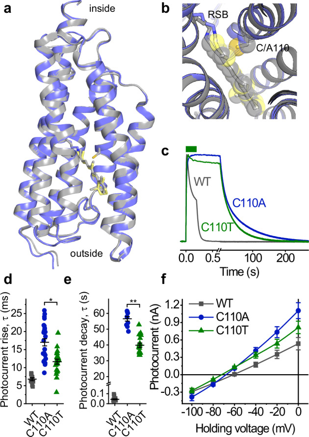
Structural information on channelrhodopsins' mechanism of light-gated ion conductance is scarce, limiting its engineering as optogenetic tools. Here, we use single-particle cryo-electron microscopy of peptidisc-incorporated protein samples to determine the structures of the slow-cycling mutant C110A of kalium channelrhodopsin 1 from Hyphochytrium catenoides (HcKCR1) in the dark and upon laser flash excitation. Upon photoisomerization of the retinal chromophore, the retinylidene Schiff base NH-bond reorients from the extracellular to the cytoplasmic side. This switch triggers a series of side chain reorientations and merges intramolecular cavities into a transmembrane K conduction pathway. Molecular dynamics simulations confirm K flux through the illuminated state but not through the resting state. The overall displacement between the closed and the open structure is small, involving mainly side chain rearrangements. Asp105 and Asp116 play a key role in K conductance. Structure-guided mutagenesis and patch-clamp analysis reveal the roles of the pathway-forming residues in channel gating and selectivity.
关于视紫红质光门控离子传导机制的结构信息匮乏,这限制了其作为光遗传学工具的工程化应用。在此,我们利用肽盘结合蛋白样品的单颗粒冷冻电子显微镜技术,来确定来自链壶菌(HcKCR1)的钾离子视紫红质1的慢循环突变体C110A在黑暗中和激光闪光激发后的结构。在视网膜发色团发生光异构化后,视黄叉席夫碱的NH键从细胞外侧重新定向到细胞质侧。这种转变引发了一系列侧链重排,并将分子内的腔合并成一条跨膜钾离子传导途径。分子动力学模拟证实钾离子通过光照状态下的通道,但不通过静息状态下的通道。关闭结构和开放结构之间的整体位移很小,主要涉及侧链重排。天冬氨酸105和天冬氨酸116在钾离子传导中起关键作用。基于结构的诱变和膜片钳分析揭示了形成通道的残基在通道门控和选择性中的作用。